. 2014 Dec;66(12):3349-58.
doi: 10.1002/art.38868.

FoxO transcription factors support oxidative stress resistance in human chondrocytes

Affiliations

FoxO transcription factors support oxidative stress resistance in human chondrocytes

Yukio Akasaki et al. Arthritis Rheumatol. 2014 Dec.

Abstract

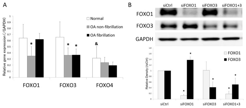
Objective: A major signaling pathway that regulates cellular aging is the insulin/insulin-like growth factor 1 (IGF-1)/phosphatidylinositol 3-kinase (PI3K)/Akt/FoxO transcription factor axis. We previously observed that FoxO transcription factors are dysregulated in aged and OA cartilage. The objective of this study was to investigate the impact of down-regulated FoxO transcription factors on chondrocytes.

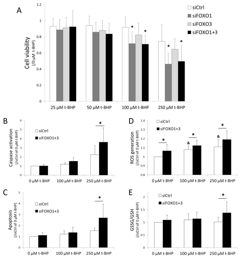
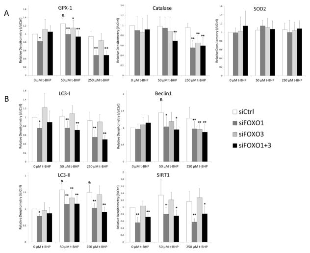
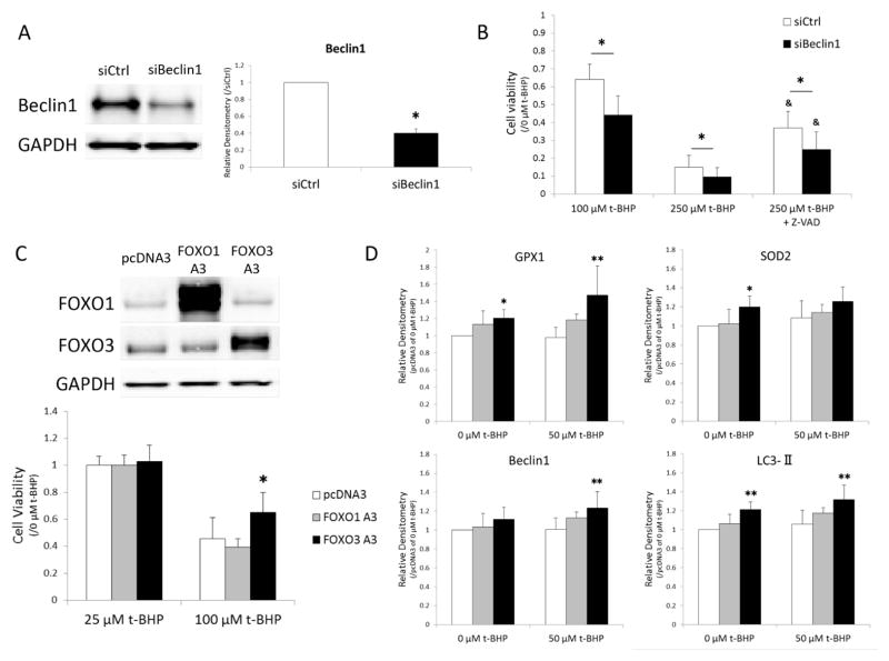
Methods: Small interfering RNAs (siRNAs) targeting FOXO1 (siFOXO1) and FOXO3 (siFOXO3) were transfected into human articular chondrocytes. Cell viability following treatment with the oxidant tert-butyl-hydroperoxide (tBHP) was measured by MTT assay. Caspase 3/7 activation and apoptotic cells were examined. Gene and protein expression of antioxidant proteins and autophagy-related proteins and changes in inflammatory mediators following treatment with interleukin-1β were assessed. Cells transfected with FOXO plasmids were also analyzed.

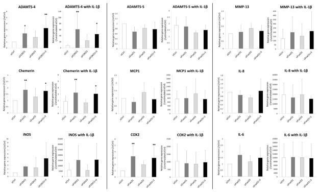
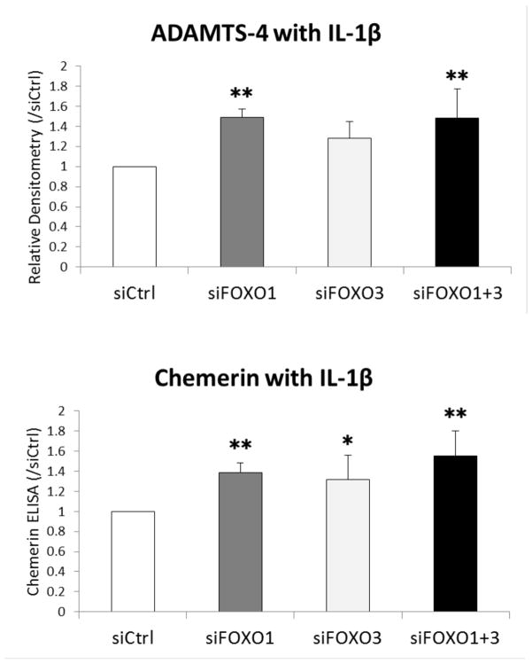
Results: Cell viability was significantly reduced by siFOXO after treatment with tBHP. Apoptosis accompanied by caspase activation was significantly increased in siFOXO-transfected chondrocytes. Knockdown of FOXO1 and FOXO1+3 resulted in significant reductions in levels of glutathione peroxidase 1 (GPX-1), catalase, light chain 3 (LC3), Beclin1, and sirtuin 1 (SIRT-1) proteins following treatment with tBHP. In contrast, the constitutive active form of FOXO3 increased cell viability while inducing GPX-1, Beclin1, and LC3 in response to tBHP. Expression and production of ADAMTS-4 and chemerin were significantly increased in siFOXO-transfected chondrocytes.

Conclusion: Reduced expression of FoxO transcription factors in chondrocytes increased susceptibility to cell death induced by oxidative stress. This was associated with reduced levels of antioxidant proteins and autophagy-related proteins. Our data provide evidence for a key role of FoxO transcription factors as regulators of chondrocyte oxidative stress resistance and tissue homeostasis.


Conflict of interest statement

The authors have no conflicts of interest.

Competing interests None.

Figures

Figure 1
Figure 1
(A) FOXO mRNA expression in human articular cartilage. RNA samples isolated from articular cartilage were analyzed by real-time PCR using primers for FOXOs as indicated. Results are from a total of 4 normal (N=4) and 5 OA donors (N=5) with separate analysis of non-fibrillated and fibrillated areas. Values are the mean and SD. * = P < 0.05 versus normal; & = P < 0.05 versus FOXO1 and FOXO3. (B) Transfection with siFOXO in human chondrocytes. Protein extracts from chondrocytes transfected with siRNAs were analyzed by western blotting using antibodies to FOXOs as indicated. Image is representative of FOXO1 and FOXO3 protein expression in siRNA transfected cells from the same samples. Graph shows the results of a total of 4 donors (N=4). Values are the mean and SD. * = P < 0.05 versus siCtrl.
Figure 2
Figure 2
(A) Changes in cell viability in siFOXO transfected chondrocytes under oxidative stress with t-BHP. Chondrocytes were transfected with siRNAs and cell viability under treatment with t-BHP was assessed by MTT assay. Cell viability was calculated as the percentage of absorbance of t-BHP treated cells (25, 50, 100, and 250 μM of t-BHP) with respect cells not treated with t-BHP (0 μM of t-BHP). Graph shows the results of a total of 8 donors (N=8). Values are the mean and SD. * = P < 0.05 versus siCtrl at each concentration of t-BHP. (B) Caspase activation and (C) induction of apoptosis in response to t-BHP in siRNA treated cells. Graphs show the results of a total of 5 donors (N=5) for caspase activation and apoptosis detection in siRNA transfected cells treated with t-BHP (100 and 250 μM). Values are the mean and SD as the ratio of t-BHP treated cells (0, 100 and 250 μM of t-BHP) with siCtrl transfected cells not treated with t-BHP (0 μM of t-BHP). * = P < 0.05 versus siCtrl at each concentration of t-BHP. (D) Changes in ROS generation and (E) redox state of glutathione in response to t-BHP in siRNA treated cells. Graphs show the results of a total of 8 donors (N=8) for ROS generation and 10 donors for the ratio of GSSG to GSH in siRNA transfected cells treated with t-BHP (100 and 250 μM). Values are the mean and SD as the ratio of t-BHP treated cells (0, 100 and 250 μM of t-BHP) with siCtrl transfected cells not treated with t-BHP (0 μM of t-BHP). * = P < 0.05 versus siCtrl at each concentration of t-BHP. & = P < 0.05 versus siCtrl at 0 μM of t-BHP.
Figure 3
Figure 3
Changes in anti-oxidant proteins and autophagy proteins in response to t-BHP in siFOXO transfected chondrocytes. Human chondrocytes transfected with siRNA were stimulated with t-BHP (50 and 250 μM) for 24 h. Protein extracts were analyzed by western blotting using antibodies to (A) anti-oxidant proteins (B) autophagy proteins and Sirt1. Graph shows the results of a total of 4 donors (N=4). Values are the mean and SD. * = P < 0.05 and ** = P < 0.01 versus siCtrl at each concentration of t-BHP. & = P < 0.05 versus siCtrl at 0 μM of t-BHP.
Figure 4
Figure 4
(A and B) The effect of Beclin1 down-regulation on cell viability in response to t-BHP. Human chondrocytes were transfected with siBeclin1 (A) and cell viability under treatment with t-BHP was examined by Resazurin assay (B). Graph shows the results of a total of 4 donors (N=4). Values are the mean and SD. * = P < 0.05 versus siCtrl. & = P < 0.05 versus siCtrl at 250 μM of t-BHP. (C and D) The effect of FOXO overexpression on cell viability and anti-oxidant and autophagy proteins. Image is representative of FOXO1 and FOXO3 protein expression in FOXO plasmid transfected cells. Cell viability under treatment with t-BHP was examined by MTT assay (C). Protein extracts were analyzed by western blotting. Graph shows the results of a total of 4 donors (N=4). Values are the mean and SD. * = P < 0.05 and ** = P < 0.01 versus pcDNA empty vector
Figure 5
Figure 5
Changes in inflammatory gene expression in response to IL-1β in siFOXO transfected chondrocytes. Human chondrocytes transfected with siRNA were stimulated with or without IL-1β (0.01 ng/ml) for 6 h. Inflammatory gene expression was analyzed by real-time PCR using primers as indicated. Graph shows the results of a total of 5 donors (N=5). Values are the mean and SD. * = P < 0.05 and ** = P < 0.01 versus siCtrl.
Figure 6
Figure 6
Changes in ADAMTS-4 and Chemerin production in response to IL-1β in siFOXO transfected chondrocytes. Human chondrocytes transfected with siRNA were stimulated with IL-1β (0.1 ng/ml) for 24 h. ADAMTS-4 protein was analyzed by western blot and Chemerin protein in the supernatant was measured by ELISA. Graph shows the results of a total of 4 donors (N=4). Values are the mean and SD. * = P < 0.05 and ** = P < 0.01 versus siCtrl.

References

    1. Lotz M, Loeser RF. Effects of aging on articular cartilage homeostasis. Bone. 2012;51:241–8. - PMC - PubMed
    1. Longo VD, Finch CE. Evolutionary medicine: from dwarf model systems to healthy centenarians? Science. 2003;299(5611):1342–6. - PubMed
    1. Lin K, Dorman JB, Rodan A, Kenyon C. daf-16: An HNF-3/forkhead family member that can function to double the life-span of Caenorhabditis elegans. Science. 1997;278(5341):1319–22. - PubMed
    1. Maiese K, Chong ZZ, Shang YC. OutFOXOing disease and disability: the therapeutic potential of targeting FoxO proteins. Trends in molecular medicine. 2008;14(5):219–27. - PMC - PubMed
    1. Rached MT, Kode A, Xu L, Yoshikawa Y, Paik JH, Depinho RA, et al. FoxO1 is a positive regulator of bone formation by favoring protein synthesis and resistance to oxidative stress in osteoblasts. Cell metabolism. 2010;11(2):147–60. - PMC - PubMed

Publication types

MeSH terms

LinkOut - more resources