. 2014 Nov 6;515(7525):138-42.
doi: 10.1038/nature13601. Epub 2014 Sep 3.

Broad and potent HIV-1 neutralization by a human antibody that binds the gp41-gp120 interface

Affiliations

Broad and potent HIV-1 neutralization by a human antibody that binds the gp41-gp120 interface

Jinghe Huang et al. Nature. .

Abstract

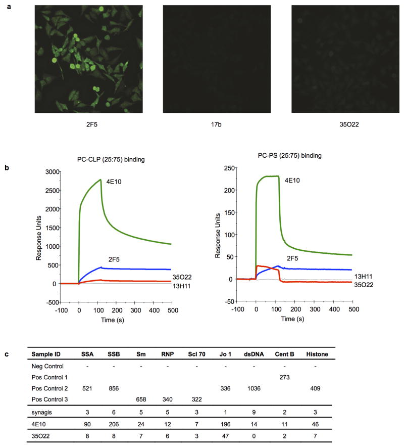
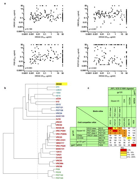
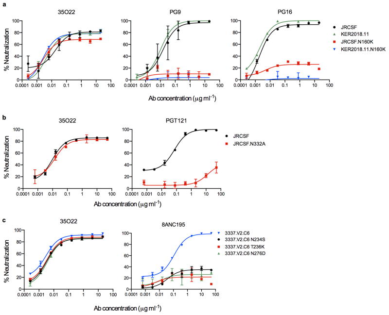
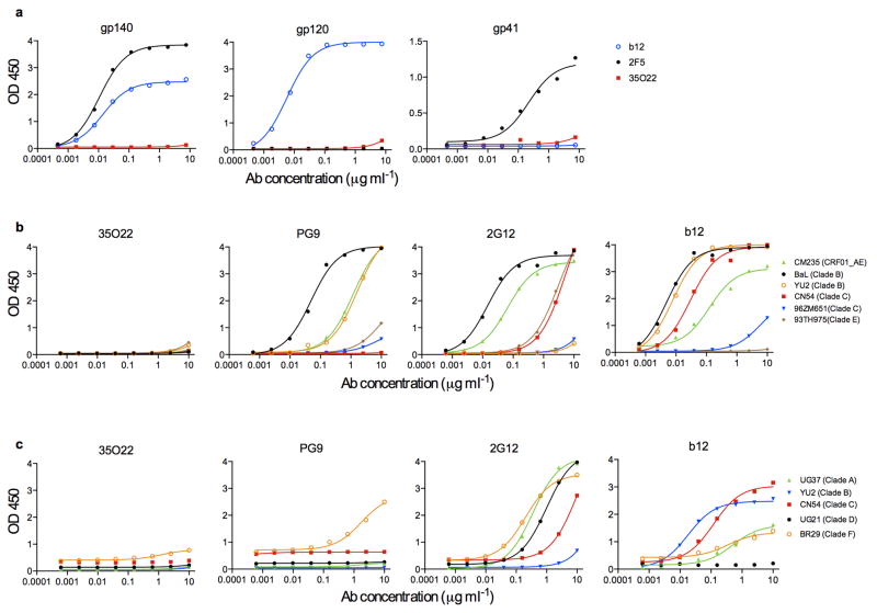
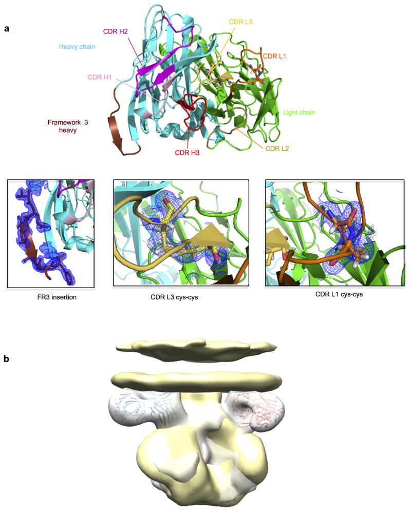
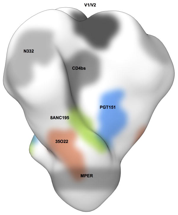
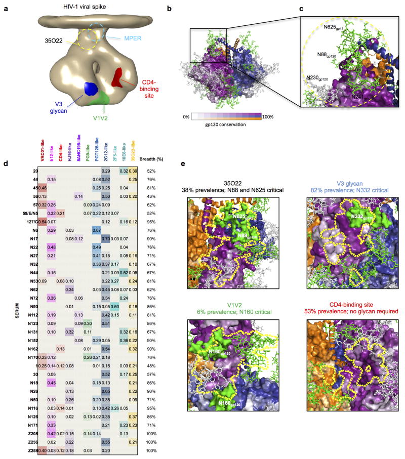
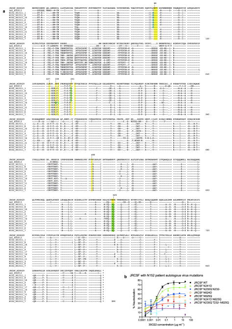
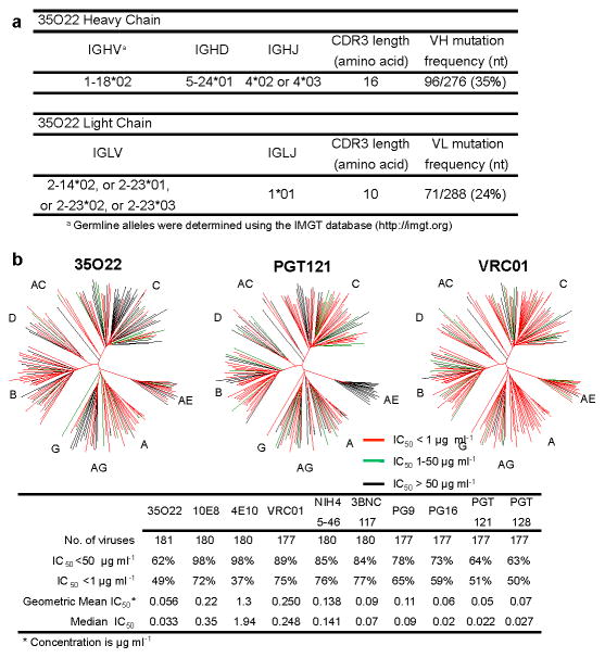
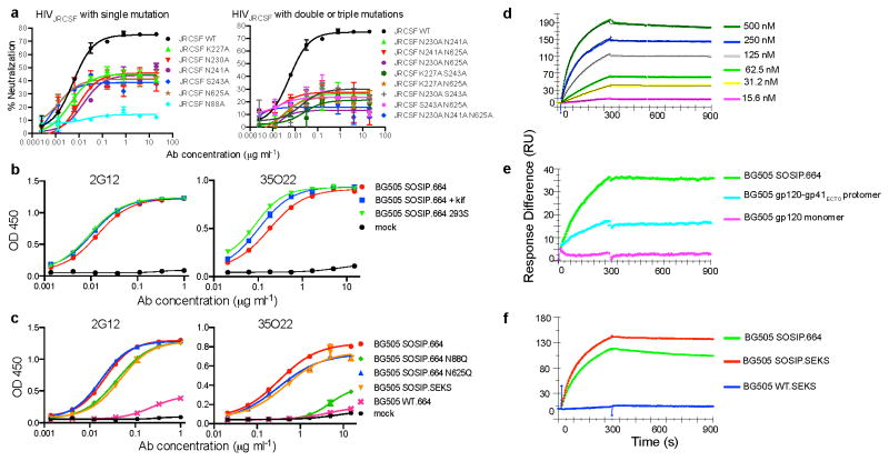
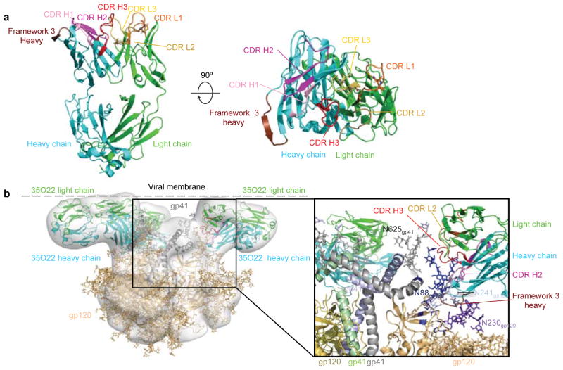
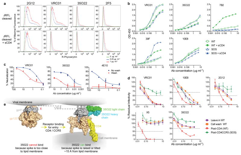
The isolation of human monoclonal antibodies is providing important insights into the specificities that underlie broad neutralization of HIV-1 (reviewed in ref. 1). Here we report a broad and extremely potent HIV-specific monoclonal antibody, termed 35O22, which binds a novel HIV-1 envelope glycoprotein (Env) epitope. 35O22 neutralized 62% of 181 pseudoviruses with a half-maximum inhibitory concentration (IC50) <50 μg ml(-1). The median IC50 of neutralized viruses was 0.033 μg ml(-1), among the most potent thus far described. 35O22 did not bind monomeric forms of Env tested, but did bind the trimeric BG505 SOSIP.664. Mutagenesis and a reconstruction by negative-stain electron microscopy of the Fab in complex with trimer revealed that it bound to a conserved epitope, which stretched across gp120 and gp41. The specificity of 35O22 represents a novel site of vulnerability on HIV Env, which serum analysis indicates to be commonly elicited by natural infection. Binding to this new site of vulnerability may thus be an important complement to current monoclonal-antibody-based approaches to immunotherapies, prophylaxis and vaccine design.


Conflict of interest statement

The authors declare no competing financial interests. Readers are welcome to comment on the online version of the paper.

Figures

Extended Data Figure 1
Extended Data Figure 1. Analysis of 35O22 autoreactivity
a, Reactivity of 35O22 with HEP-2 epithelial cells. 2F5 was used as a positive control and 17b as a negative control. Antibody concentration was 25 μg/ml. All pictures are shown at 400x magnification. b, SPR analysis of 35O22 binding to anionic phospholipids. 35O22 was injected over PC-CLP liposomes or PC-PS liposomes immobilized on the BIAcore L1 sensor chip. 4E10 and 2F5 were used as positive controls and 13H1 as a negative control. c, Reactivity of 35O22 with autoantigens detected in Luminex assay. 4E10 was used as a positive control. Synagis, an anti-RSV monoclonal antibody, was used as a negative control. SSA, Sjogren’s syndrome antigen A; SSB, Sjogren syndrome antigen B; Sm, Smith antigen; RNP, ribonucleoprotein; Scl 70, scleroderma 70; Jo1, antigen; CentrB, centromere B. A positive response is >120 units.
Extended Data Figure 2
Extended Data Figure 2. Neutralization similarities between 35O22 and other HIV-1 bNAbs
a, Correlation (Spearman) between the neutralization potencies of 35O22 and the indicated antibody against 172 pseudoviruses. Representatives from all four major sites of vulnerability are shown. Resistant strains corresponding to values of >50 μg/ml are plotted as 50. b, Neutralization-based clustering of bNabs over a set of 172 diverse HIV-1 strains. A putative epitope-specific clustering cutoff is shown as a dashed line. Antibodies are colored according to the respective target site of vulnerability: red (CD4bs), blue (glycan-V3), V1V2 (green), light blue (MPER), and other (purple). 35O22 (yellow) clusters separately from all other antibodies, indicating a novel mechanism of neutralization. c, 35O22 competition with other bNAbs on HIVJRFL VLPs with the trimer stabilizing SOS mutations in an ELISA assay. Biotin-bNAbs were titrated into the ELISA at increasing concentrations in the presence of excess (10 μg ml−1) cold competitor neutralizing antibodies. Values in the table indicates percentage binding of biotin-nAbs in the presence of cold-competitor. ND = not done
Extended Data Figure 3
Extended Data Figure 3. 35O22 binds to N-linked glycans
a, Neutralization by 35O22 plateaus below 80% against several pseudoviruses. b, Neutralization activity of mAbs against JRCSF pseudoviruses generated in the presence of glycosidase-inhibitors, such as kifunensine (25 μM), NB-DNJ (500 μM) or swainsonine (20 μM). Error bars denote one standard error of the mean (s.e.m.).
Extended Data Figure 4
Extended Data Figure 4. Neutralization of 35O22 against pseudovirus mutants known to knock out activity against known glycan-specific antibodies
a, Neutralization of 35O22 against JRCSF or KER2018.11 with or without the N160K mutation. PG9 and PG16 were used as positive controls. b, Neutralization of 35O22 against N332A mutants of JRCSF. PGT121 was used as a positive control. c, Neutralization of 35O22 against N234S, T236K and N276D mutants of 3337.V2.C6. 8ANC195 was used as a positive control. Error bars denote one standard error of the mean (s.e.m.).
Extended Data Figure 5
Extended Data Figure 5. Binding specificity of 35O22
ELISA binding of indicated mAbs to HIVYU2 gp140 foldon trimer, gp120, and gp41 monomers (a). ELISA binding of gp120 (b) and gp140 (c) monomers from different HIV-1 subtypes.
Extended Data Figure 6
Extended Data Figure 6. 35022 Fab features
a. 35022 is seen looking down on the combining site from the viewpoint of antigen in ribbons with the CDR colored as in Figure 3a. Insets show structural details of the framework 3 insertion, disulfides in CDR L1 and CDR L3 with electron density 2F0−Fc contoured at 1σ.Location of the viral membrane. b. Superposition of BaL gp160 negative stain (yellow surface) with the negative stain reconstruction of soluble BG505 SOSIP in complex with 35O22 (grey surface) gives an estimation of the viral membrane location relative to 35O22 antibody as shown in Figure 3b.
Extended Data Figure 7
Binding site of 35O22 on the HIV Env trimer. Binding site of 35O22 (red) relative to those of PGT151 (blue) or 8ANC195 (green) are shown.
Extended Data Figure 8
Extended Data Figure 8. A new site of HIV-1 vulnerability at the interface of gp120 and gp41 and prevalence of targeting
a, Dominant sites of vulnerability to neutralizing antibody elicited by natural infection, shown in the context of an EM tomogram from the BAL viral spike. The viral membrane is positioned at the top of the spike. It is unclear if 35O22 and MPER antibodies bind to this form of the viral spike, and approximate locations for these are shown in dotted outlines. b, Viral spike from the soluble BG505 SOSIP context, shown in the same orientation as a, with gp120 surface colored by conservation from 0–100%, from 4265 HIV-1 strains (white to purple for protomer 1 with scale shown, white to blue for protomer 2 and white to orange for protomer 3), with glycans shown in green when present in more than 90% of strains, in grey when present in 30–90% of strains and not shown otherwise. c, 35O22-identified site of HIV-1 vulnerability, comprises both conserved amino acids and a cluster of glycans, including N88 from gp120 and N625 from gp41. N230 and N241 are not present in BG505 strain. The 35O22 epitope is shown in yellow dotted line. d, Neutralization fingerprints for 35O22 and for antibodies encompassing ten different epitope specificities representing the other four known major sites of Env vulnerability were used to interrogate the serum specificities of 34 HIV-infected patients. Values (with proportional color intensities) predict the fraction of serum neutralization that can be attributed to each antibody specificity. Possible 35O22-like signals were predicted for 13 of the sera (values >0.2), while strong signals were observed in 3 of the sera (values >0.3). A panel of 21 HIV-1 strains was used in the neutralization analysis and for computing serum breadth. e, Sites of HIV-1 vulnerability to neutralizing antibody outlined by a yellow line. Prevalence in 34-donor cohort indicated along with critical glycans.
Extended Data Figure 9
Extended Data Figure 9. Autologous virus Env sequence and the impact of variants on 35O22 neutralization
a, A total of 12 single-genome amplicons from plasma of patient N152 were sequenced. Donor Env sequences together with the reference sequences of JRCSF and LAI are aligned. Amino acids critical for 35O22 neutralization of JRCSF and LAI are labeled in yellow. Differences between autologous and JRCSF sequences are labeled in green. b, 35O22 neutralization of JRCSF pseudovirus or variants containing the autologous virus mutations from patient N152. Error bars denote one standard error of the mean.
Figure 1
Figure 1. Analyses of 35O22 sequence and neutralization
a, Inferred germline genes encoding the variable regions 35O22. b, Neutralizing activity of antibodies against a 181-isolate Env-pseudovirus panel. Dendrograms indicate the gp160 protein distance of HIV-1 primary isolate Env glycoproteins. Data below the dendrogram show the number of tested viruses, the percentage of viruses neutralized and the geometric mean or median IC50 for viruses neutralized with an IC50 < 50 μg ml−1.
Figure 2
Figure 2. Binding specificity of 35O22
a, Neutralization of HIVJRCSF pseudovirus or variants containing the indicated mutations. b, Binding to BG505 SOSIP.664 trimer produced in cells treated with kifunensine or deficient in glycan processing (293S). c, Binding to BG505 trimers with the indicated mutations. BG505 SOSIP.SEKS lacks the furin cleavage site. BG505 WT.664 lacks stabilizing mutations and the antigen primarily represents gp41. d, SPR analysis of binding to immobilized BG505 SOSIP.664 trimers. e, Binding of 35022 (250 nM) to BG505 SOSIP.664 trimers, gp120-gp41ECTO protomers, or monomeric gp120. f, Binding to BG505 SOSIP.664, BG505 SOSIP.SEKS, or the BG505 WT.SEKS lacking the SOSIP mutations.
Figure 3
Figure 3. Structure of Fab 35O22 and EM reconstruction of complex with HIV-1 Env
a, Cartoon representation (left) of unbound 35O22 Fab. Heavy chain is in cyan, light chain in green, and the heavy chain FR3 insertion in chocolate. (right) 90° rotation, looking down on combining site. b, EM reconstruction of BG505 SOSIP.664 in complex with 35O22 Fab, with fitted crystal structures. gp120 (light orange) and gp41 (grey) shown with 35O22 (as in a). The approximate location of the viral membrane is indicated. Glycans N241 and N230 are not part of the BG505 sequence, but have been modelled for reference.
Figure 4
Figure 4. Binding or neutralization in the context of a lipid membrane
a, Staining of cell surface expressed HIVJRFL Env. b, ELISA assay of antibody binding to WT or SOS HIVJRFL VLPs. The CD4-inducible 39F antibody or gp41-specific 7B2 are used as controls. c, Access to the HIVJRFL Env trimer on pseudovirions based upon washing the antibody-pseudovirion mixture prior to infecting cells. d, Kinetic assay of HIVJRFL neutralization. See Methods for a description of individual formats. e, Schematic of conformational change resulting in raising of the trimer spike required to permit access of 35O22 to its epitope.

References

    1. Kwong PD, Mascola JR. Human antibodies that neutralize HIV-1: identification, structures, and B cell ontogenies. Immunity. 2012;37:412–425. - PMC - PubMed
    1. Doria-Rose NA, et al. Breadth of human immunodeficiency virus-specific neutralizing activity in sera: clustering analysis and association with clinical variables. J Virol. 2010;84:1631–1636. - PMC - PubMed
    1. Sather DN, et al. Factors associated with the development of cross-reactive neutralizing antibodies during human immunodeficiency virus type 1 infection. J Virol. 2009;83:757–769. - PMC - PubMed
    1. Walker LM, et al. A limited number of antibody specificities mediate broad and potent serum neutralization in selected HIV-1 infected individuals. PLoS Pathog. 2010;6:e1001028. - PMC - PubMed
    1. Simek MD, et al. Human immunodeficiency virus type 1 elite neutralizers: individuals with broad and potent neutralizing activity identified by using a high-throughput neutralization assay together with an analytical selection algorithm. J Virol. 2009;83:7337–7348. - PMC - PubMed

Publication types

MeSH terms

Associated data