. 2014 Sep 3;34(36):12001-14.
doi: 10.1523/JNEUROSCI.1663-14.2014.

Thalamic NMDA receptor function is necessary for patterning of the thalamocortical somatosensory map and for sensorimotor behaviors

Affiliations

Thalamic NMDA receptor function is necessary for patterning of the thalamocortical somatosensory map and for sensorimotor behaviors

Hiroyuki Arakawa et al. J Neurosci. .

Abstract

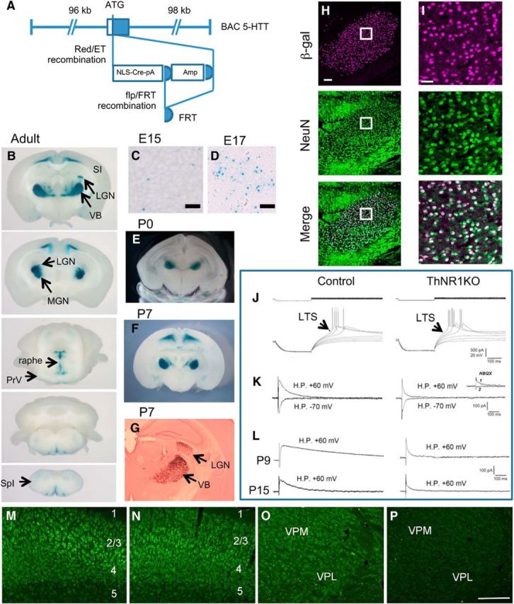
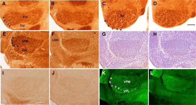
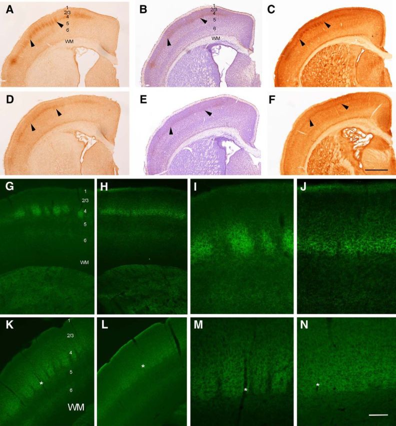
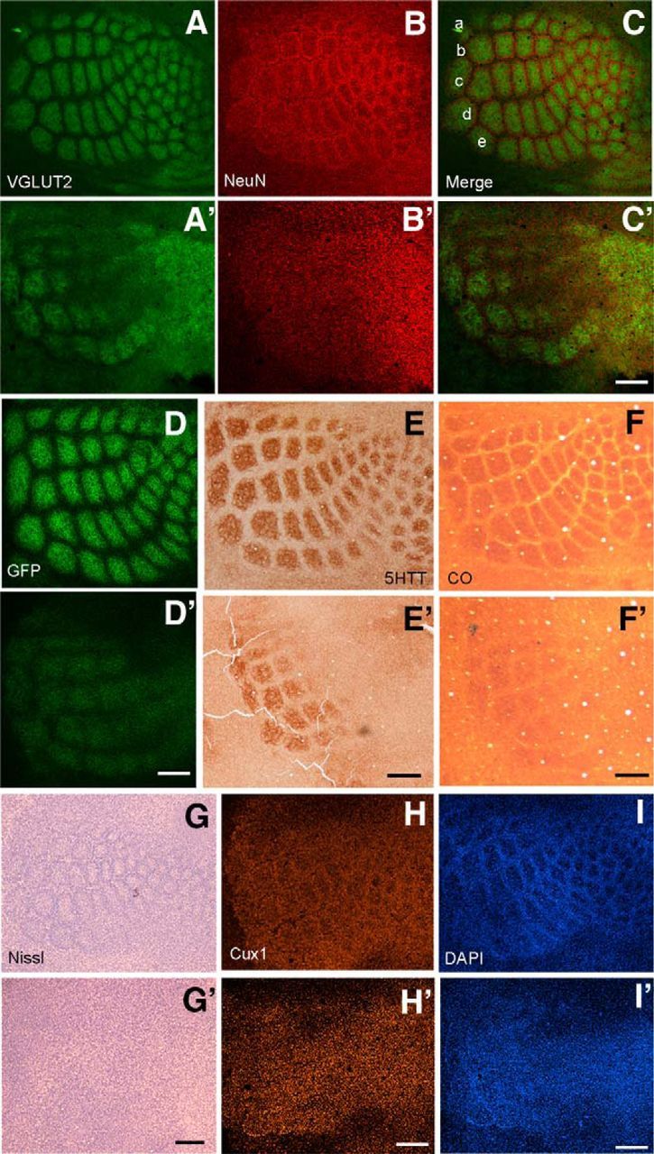
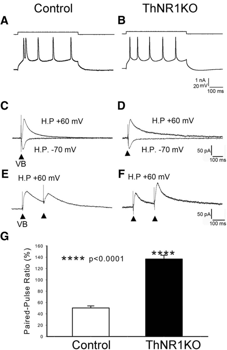
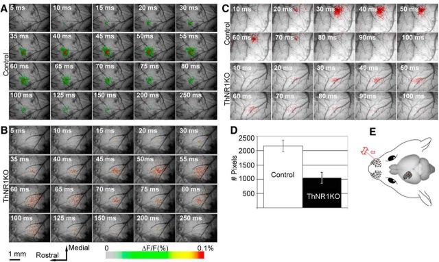
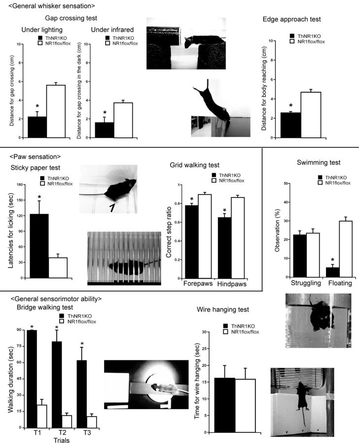
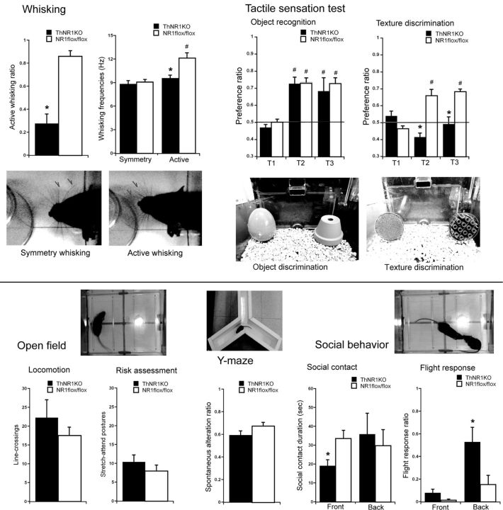
NMDARs play a major role in patterning of topographic sensory maps in the brain. Genetic knock-out of the essential subunit of NMDARs in excitatory cortical neurons prevents whisker-specific neural pattern formation in the barrel cortex. To determine the role of NMDARs en route to the cortex, we generated sensory thalamus-specific NR1 (Grin1)-null mice (ThNR1KO). A multipronged approach, using histology, electrophysiology, optical imaging, and behavioral testing revealed that, in these mice, whisker patterns develop in the trigeminal brainstem but do not develop in the somatosensory thalamus. Subsequently, there is no barrel formation in the neocortex yet a partial afferent patterning develops. Whisker stimulation evokes weak cortical activity and presynaptic neurotransmitter release probability is also affected. We found several behavioral deficits in tasks, ranging from sensorimotor to social and cognitive. Collectively, these results show that thalamic NMDARs play a critical role in the patterning of the somatosensory thalamic and cortical maps and their impairment may lead to pronounced behavioral defects.

Keywords: barrel cortex imaging; barreloids; barrels; glutamate; pattern formation.


Figures

Figure 1.
Figure 1.
Generation of 5-HTTCre Tg and ThNR1KO mice. A, The translational initiation site of the serotonin transporter (5-HTT) gene on a BAC clone was replaced with the coding sequence of NLS-Cre, and the Amp selection marker was subsequently removed by flp/FRT recombination in bacteria. This BAC construct was microinjected into fertilized eggs to generate 5-HTTCre Tg mice. B, In 5-HTTCre Tg mice crossed with RNZ reporter mice, Cre-mediated recombination was detected in the sensory thalamus such as VB (somatosensory), lateral geniculate nucleus (LGN; visual) and medial geniculate nucleus (MGN; auditory), raphé, and deep layers of medial parts of the neocortex, but was not detected in the SI cortex and the trigeminal brainstem such as the PrV and SpI. LacZ-stained images of a 1-mm-thick coronal slice of 5-HTTCre Tg208;RNZ mouse at P38. C, D, Cre-meditated recombination assessed by LacZ stain in VB was first detected at E15 (C) and was dense at E17 (D). Scale bars: 25 μm. 5-HTTCre;RNZ embryo 30-μm-thick coronal slices. E, F, In early postnatal development, Cre-mediated recombination in VB is very high. Coronal slices of 5-HTTCre;RNZ mice at P0 (E; 400-μm-thick slice with LacZ stain) and P7 (F; 1-mm-thick slice with LacZ stain). G, A 20-μm-thick coronal section from a P7 mouse (LacZ/Neutral red stain). H, I, Double immunohistochemistry of VB of 5-HTTCre;RNZ mouse slice (30 μm thick) at P5 with anti-β-galactosidase and anti-NeuN (a neuronal nuclear marker) antibodies. I, Higher magnification images of the boxed regions in H. These images were used for quantification of Cre-mediated recombination. Scale bars: H, 100 μm; I, 25 μm. JL, Comparison of electrophysiological properties of VB neurons between ThNR1KO and control mice. J, VB cells show similar low-threshold Ca2+ spikes. K, Different duration in EPSCs at +60 mV. The inset shows that the EPSC at +60 mV of KO VB is completely blocked by NBQX (trace 1 before vs trace 2 after NBQX), indicating absence of NMDAR-mediated response. L, Differences in developmental changes in decay of EPSCs at +60 mV. All of these confirm that VB cells in ThNR1KO mice lack NMDAR-mediated postsynaptic responses. M, N, NR1 expression in the barrel cortex of a control (M) and a ThNR1KO adult mice (N). Note that there is no apparent difference in cytoplasmic staining of cortical neurons between the control and knock-out cases. Cortical layers are indicated. O, P, In control mice neuron in the VPM and VPL components of the VB also stain positive for NR1 (O), but staining is absent in the ThNR1KO VPM and VPL (P), even though some distinct, unbound fluorescent secondary antibody particles can be seen. Scale bar, M–P, 100 μm.
Figure 2.
Figure 2.
Whisker-specific patterning in subcortical trigeminal centers. A–D, Barrelette patterns in the PrV (A, B) and SpI (C, D) of the spinal trigeminal nucleus in ThNR1KO mice at P5 (B, D) appear very similar to those of P5 controls (A, C). CO staining, barrelette rows a–e are indicated. Unlike the trigeminal brainstem, barreloids are absent in the VPM and VPL components of the VB in ThNR1KO mice (F, H, J, L). Photomicrographs in E–H compare the VB of ThNR1KO and control mice at P5, at P7 (I, J), and at P9 (K, L) with CO staining (E, F), Nissl staining (G, H), 5-HTT (I, J), and VGLUT2 immunohistochemistry (K, L). Scale bar, 200 μm. LGN, Lateral geniculate nucleus; TrV, trigeminal tract.
Figure 3.
Figure 3.
Cortical lamination and thalamocortical axon targeting. Immunolabeling for 5-HTT (A, D), Nissl staining (B, E), and CO histochemistry (C, F) in coronal sections indicate that TCAs in ThNR1KO mice target primarily layer 4 (D–F) but, unlike the controls (A–C), they do not form discrete terminal patches and cellular barrels are absent. G–J, Show VGLUT2 immunostaining at P7 in control (G, I) and ThNR1KO mice (H, J) at low (G, H) and high (I, J) power. Note that the apparent density of TCAs in the knock-out case is weaker. K–N, Illustrate VGLUT2 immunostaining in adult control (K, M) and ThNR1KO mice (L, N). L and N are higher power views of layer 4 and asterisks mark the same blood vessels in low- and high-power micrographs. Cortical layers are marked in some photomicrographs. WM, white matter. Arrowheads point to layer 4 barrel fields. Scale bars: (in F) A–F, 400 μm; (in N) I, J, M, N, 50 μm; G, H, K, L, 150 μm.
Figure 4.
Figure 4.
Altered barrel fields in ThNR1KO mice. A variety of cellular, axonal, and mitochondrial markers all showed that barrels fail to develop in ThNR1KO mice even though an incomplete and ill-defined TCA terminal patterning related to large, caudal whiskers developed. A, B, Barrel patterning as seen with VGLUT2 (A) and NeuN (B) of control mice. A′, B′, Show similar cortical views from ThNR1KO mice. C, C′, Respective merged images; whisker barrel rows a–e are indicated. D, D′, Compare TCA terminal patterning in control (TCA-GFP) and ThNR1KO;TCA-GFP mice at P7, respectively, visualized by GFP fluorescence. Note that with similar imaging conditions the GFP labeling is weak in the mutant (ThNR1KO;TCA-GFP) mouse cortex. Similar pairs of micrographs are shown for 5-HTT immunostaining (E, E′), CO staining (F, F′), Nissl staining (G, G′), Cux1 immunostaining (H, H′), and DAPI staining (I, I′). Scale bars, 250 μm.
Figure 5.
Figure 5.
PPR in control and ThNR1KO mice. A, B, In both control and ThNR1KO mice, layer 4 cells showed adapting regular spiking during membrane depolarization. C, D, Stimulation of VB induced EPSCs with a long duration at +60 mV holding potential (H.P.) and with a short duration at −70 mV not only in control, but also in ThNR1KO mice, indicating that layer 4 cells have both AMPARs and NMDARs. E, In control mice, the amplitude of the second EPSC was smaller than that of the first EPSC (paired-pulse depression), indicating that the TCAs have a high transmitter release probability. Arrowheads indicate VB stimulation. The averaged PPR was 50.4 ± 3.7% (n = 15; G, white bar). F, In ThNR1KO mice, the second EPSC was always larger than the first EPSC (paired-pulse facilitation), indicating that the transmitter release probability of thalamocortical terminals was low. The averaged PPR was 136.8 ± 6.7% (n = 9, G, black bar). G, The difference in PPR between control and ThNR1KO mice was highly significant (p < 0.0001).
Figure 6.
Figure 6.
Comparison of voltage-sensitive dye optical imaging in control and ThNR1KO barrel cortex. A, Pseudocolor maps showing single-whisker (E2) stimulation fluorescence changes in a control mouse. The time after stimulus onset is indicated at the top left corner of each image; duration of each frame is 5 ms. B, Voltage-sensitive dye optical images showing single-whisker stimulation fluorescence changes in a ThNR1KO mouse. The time after stimulus onset is indicated at the top left corner of each image; duration of each frame is 5 ms. Note that the activated area in the barrel cortex of the knock-out mouse has a much less distinct border and is less focalized compared with the control case. C, Examples of imaging data showing only the pixels (red dots) with the highest value (>90% of all pixels in the entire field of view) of fluorescence changes after C2 whisker stimulation in a control and a ThNR1KO mouse. The time after stimulus onset is indicated at the top left corner of each image; duration of each frame is 5 ms. D, Quantitative analysis of the number of pixels with overthreshold value of signal (x-axis) for the control and ThNR1KO mice (y-axis). Number of pixels with the highest value of fluorescence is calculated at 40 ms after the stimulus onset. E, Illustration of the C2 whisker stimulation and the barrel field.
Figure 7.
Figure 7.
Whisker and paw sensation is impaired in ThNR1KO mice. In the gap-crossing test, ThNR1KO mice cross shorter gap distances than controls (NR1flox/flox) both in the light (t(14) = 5.73782, p < 0.001) and in the dark (infrared) condition (t(10) = 3.4295, p = 0.0063). In the edge-approach test, ThNR1KO mice reach a shorter distance compared with control mice (t(14) = 8.6346, p < 0.0001). In the swimming test, ThNR1KO mice show a similar level of paddling (t(14) = 0.2614, p = 0.797), but significantly shorter floating time than controls (t(14) = 8.8859, p < 0.0001). ThNR1KO mice display a longer latency for licking a sticky paper off their hindpaw than controls (t(8) = 3.231, p = 0.012). In the grid-walking test, ThNR1KO mice show lower correct step ratio than controls for both the forepaws (t(14) = 4.2037, p = 0.0009) and the hindpaws (t(14) = 4.62988, p = 0.0004). In the bridge-walking test, ThNR1KO mice show significantly longer duration for reaching a platform than controls (two-way ANOVA: strain F(1,14) = 81.616, p < 0.0001; trial F(2,28) = 5.141, p = 0.0125; and strain × trial F(2,28) = 1.373, p = 0.2699). However, in the wire hanging test, there was no significant difference in hanging duration (t(14) = 0.0435, p = 0.9659) between the ThNR1KO and the control mice.
Figure 8.
Figure 8.
Whisking and tactile sensation are impaired in ThNR1KO mice. ThNR1KO mice show lower ratio of active whisking when the whiskers contact an object compared with control (NR1flox/flox) mice (t(14) = 6.2515, p < 0.001). This ratio was calculated by the number of active whisking observed during the total whisker contacts with an object (at least 10 events for each mouse). ThNR1KO mice display similar frequencies of whisker vibration during symmetry whisking, but lower frequencies during active whisking compared with controls (two-way ANOVA: strain F(1,27) = 6.976, p = 0.0136; trial F(1,27) = 0.646, p = 0.4287; and strain × trial F(1,27) = 5.079, p = 0.0325). In the object-recognition test, there were no significant differences between the knock-out and control groups throughout trials. However, the preference ratios on Trials 2 and 3 were higher than Trial 1 (two-way ANOVA: strain F(1,14) = 0.880, p = 0.3641; trial F(2,28) = 20.509, p < 0.001; and strain × trial F(2,28) = 0.099, p = 0.9062). In the texture-discrimination test, ThNR1KO mice show significantly lower preference than controls on Trials 2 and 3 but not on Trial 1 (two-way ANOVA: strain F(1,14) = 19.615, p = 0.0006; trial F(2,28) = 5.258, p = 0.0115; and strain × trial F(2,28) = 20.628, p < 0.0001). In the open-field test, there were no significant differences in locomotion (t(10) = 0.89204, p = 0.3933) or risk assessment (t(14) = 0.9876, p = 0.3401). In the Y-maze (spatial ability), there was no group difference in spontaneous alteration (t(14) = 1.6723, p = 0.1166). However, in the social behavior test, ThNR1KO mice display lower contacts toward the front of the opponent (t(14) = 2.5903, p = 0.021), while similar contacts toward the back of the opponent (t(14) = 0.4318, p = 0.6725). ThNR1KO mice also show higher flight response ratio when approached by another mouse from the back (t(14) = 2.492, p = 0.0259), but not so much when approached from the front (t(8) = 1.9151, p = 0.0918).

References

    1. Agmon A, Connors BW. Correlation between intrinsic firing patterns and thalamocortical synaptic responses of neurons in mouse barrel cortex. J Neurosci. 1992;12:319–329. - PMC - PubMed
    1. Ahissar E, Knutsen PM. Object localization with whiskers. Biol Cybern. 2008;98:449–458. doi: 10.1007/s00422-008-0214-4. - DOI - PubMed
    1. Arsenault D, Zhang ZW. Developmental remodelling of the lemniscal synapse in the ventral basal thalamus of the mouse. J Physiol. 2006;573:121–132. doi: 10.1113/jphysiol.2006.106542. - DOI - PMC - PubMed
    1. Barnéoud P, Gyger M, Andrés F, van der Loos H. Vibrissa-related behavior in mice: transient effect of ablation of the barrel cortex. Behav Brain Res. 1991;44:87–99. doi: 10.1016/S0166-4328(05)80242-6. - DOI - PubMed
    1. Brasier DJ, Feldman DE. Synapse-specific expression of functional presynaptic NMDA receptors in rat somatosensory cortex. J Neurosci. 2008;28:2199–2211. doi: 10.1523/JNEUROSCI.3915-07.2008. - DOI - PMC - PubMed

Publication types

MeSH terms

Substances

LinkOut - more resources