. 2014 Sep 5:11:153.
doi: 10.1186/s12974-014-0153-z.

The immune response after hypoxia-ischemia in a mouse model of preterm brain injury

The immune response after hypoxia-ischemia in a mouse model of preterm brain injury

Anna-Maj Albertsson et al. J Neuroinflammation. .

Abstract

Background: Preterm brain injury consists primarily of periventricular leukomalacia accompanied by elements of gray-matter injury, and these injuries are associated with cerebral palsy and cognitive impairments. Inflammation is believed to be an important contributing factor to these injuries. The aim of this study was to examine the immune response in a postnatal day (PND) 5 mouse model of preterm brain injury induced by hypoxia-ischemia (HI) that is characterized by focal white and gray-matter injury.

Methods: C57Bl/6 mice at PND 5 were subjected to unilateral HI induced by left carotid artery ligation and subsequent exposure to 10% O2 for 50 minutes, 70 minutes, or 80 minutes. At seven days post-HI, the white/gray-matter injury was examined. The immune responses in the brain after HI were examined at different time points after HI using RT-PCR and immunohistochemical staining.

Results: HI for 70 minutes in PND 5 mice induced local white-matter injury with focal cortical injury and hippocampal atrophy, features that are similar to those seen in preterm brain injury in human infants. HI for 50 minutes resulted in a small percentage of animals being injured, and HI for 80 minutes produced extensive infarction in multiple brain areas. Various immune responses, including changes in transcription factors and cytokines that are associated with a T-helper (Th)1/Th17-type response, an increased number of CD4+ T-cells, and elevated levels of triggering receptor expressed on myeloid cells 2 (TREM-2) and its adaptor protein DNAX activation protein of 12 kDa (DAP12) were observed using the HI 70 minute preterm brain injury model.

Conclusions: We have established a reproducible model of HI in PND 5 mice that produces consistent local white/gray-matter brain damage that is relevant to preterm brain injury in human infants. This model provides a useful tool for studying preterm brain injury. Both innate and adaptive immune responses are observed after HI, and these show a strong pro-inflammatory Th1/Th17-type bias. Such findings provide a critical foundation for future studies on the mechanism of preterm brain injury and suggest that blocking the Th1/Th17-type immune response might provide neuroprotection after preterm brain injury.


Figures
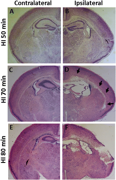
Figure 1
Figure 1
Representative photomicrographs of thionin/acid fuchsin-stained brain sections. It shows different degrees of injury in the ipsilateral hemisphere at 3 days after HI with 50 minutes (A, B, n = 12), 70 minutes (C, D, n = 14), or 80 minutes (E, F, n = 16) of hypoxia. Arrows in (D) indicate focal subcortical white-matter injury in the ipsilateral hemisphere of the HI 70-minute mouse brain.
Figure 2
Figure 2
Brain injury after 50-minute hypoxia-ischemia (HI) insult. Dot graphs show the volume difference between the ipsilateral and contralateral hemispheres in gray matter (A) and white matter (B) at 7 days after 50-minute HI in PND 5 mice (n = 16). Abbreviations: Contra: contralateral hemisphere; Ipsi: ipsilateral hemisphere. *P < 0.05 by Student’s unpaired t-test. Data are presented as mean ± SEM. (C-H) Representative photomicrographs of thionin/acid fuchsin-stained sections show diffuse injury in the subcortical white matter area (D and G) and focal granular layer loss in the hippocampus CA3 area (D and H) that is only observed in the ipsilateral hemisphere and not in the contralateral hemisphere (C, E, and F) at 3 days after HI in PND 5 mice. Arrows in (D) indicate focal injury in the CA1 granular layer of the hippocampus in the ipsilateral hemisphere.
Figure 3
Figure 3
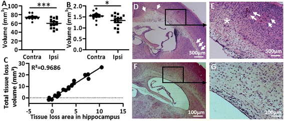
Brain injury after 70-minute hypoxia-ischemia (HI) insult. Dot graphs showing the volume difference between the contralateral and ipsilateral hemispheres in gray matter (A) and white matter (B) at 7 days after 70-minute HI in PND 5 mice (n = 13). *P < 0.05, ***P < 0.001 using Student’s unpaired t-test. Data are presented as mean ± SEM. Abbreviations: Contra: contralateral hemisphere; Ipsi: ipsilateral hemisphere. (C) The simple linear regression analysis shows the linear correlation between the two evaluation methods. (D-G) Representative photomicrographs of H&E-stained sections show focal injury in the subcortical white matter in the ipsilateral hemispheres at 3 days after HI in PND 5 mice (D and E) but not in PND 9 mice (F and G) with a similar degree of injury. Arrows in (D) show subcortical white-matter injury. Arrows and star in (E) show focal gray-matter injury in the ipsilateral hemisphere of the PND 5 HI mouse brain.
Figure 4
Figure 4
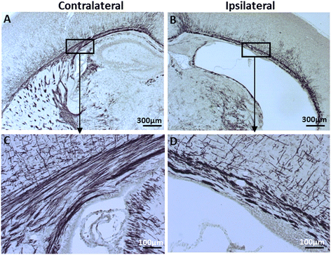
Impaired myelin structure at 7 days after 70-minute hypoxia-ischemia (HI). Representative photomicrographs of myelin basic protein (MBP)-stained brain sections at the hippocampal level show impaired myelin development and disrupted myelin structure in the subcortical white matter of the ipsilateral hemisphere (B and D) compared with the contralateral hemisphere (A and C).
Figure 5
Figure 5
TREM-2 expression after 70-minute hypoxia-ischemia (HI) insult. Representative photomicrographs show TREM-2+ immunohistochemical staining in the contralateral (A) and ipsilateral (B) hemispheres at 24 hours after HI. (C and D) TREM-2+ cells in the hippocampus in the brain parenchyma (C) and blood vessels (D). (E) TREM-2+ cells in the choroid plexus at 24 hours after HI. (F) A higher magnification of TREM-2+ staining. (G-L) TREM-2 staining (H) co-localizes (G, arrows) with microglia marker Iba-1 (I) in the choroid plexus at 24 hours after HI. (J) DAPI nuclear staining.
Figure 6
Figure 6
DAP12 expression after a 70-minute hypoxia-ischemia (HI) insult. Representative photomicrographs show expression of DAP12 (green) and its co-localization with the microglia marker Iba-1 (red) after HI. DAP12 and Iba-1 double-positive cells were found in the dentate gyrus (A-D) at 24 hours after HI, and in the proximity of blood vessels in the CA1 area of the hippocampus (E-H) at 3 days after HI.
Figure 7
Figure 7
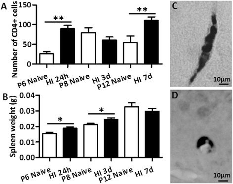
CD4 + T-cells in the mouse brain after hypoxia-ischemia (HI) in postnatal day (PND) 5 mice. The total number of CD4+ cells in blood vessels and in tissue from both of the brain hemispheres from a section at the hippocampus level (A) and spleen weight (B) in uninjured control (Cont) mice versus HI mice at different time points after HI. *P < 0.05, **P < 0.01, ***P < 0.001 using Student’s unpaired t-test. Data are presented as mean ± SEM. (C and D) Representative immunostainings show CD4+ cells in blood vessels in the ipsilateral subcortical white matter (C) and ipsilateral hippocampus CA1 area (D) at seven days post-HI.
Figure 8
Figure 8
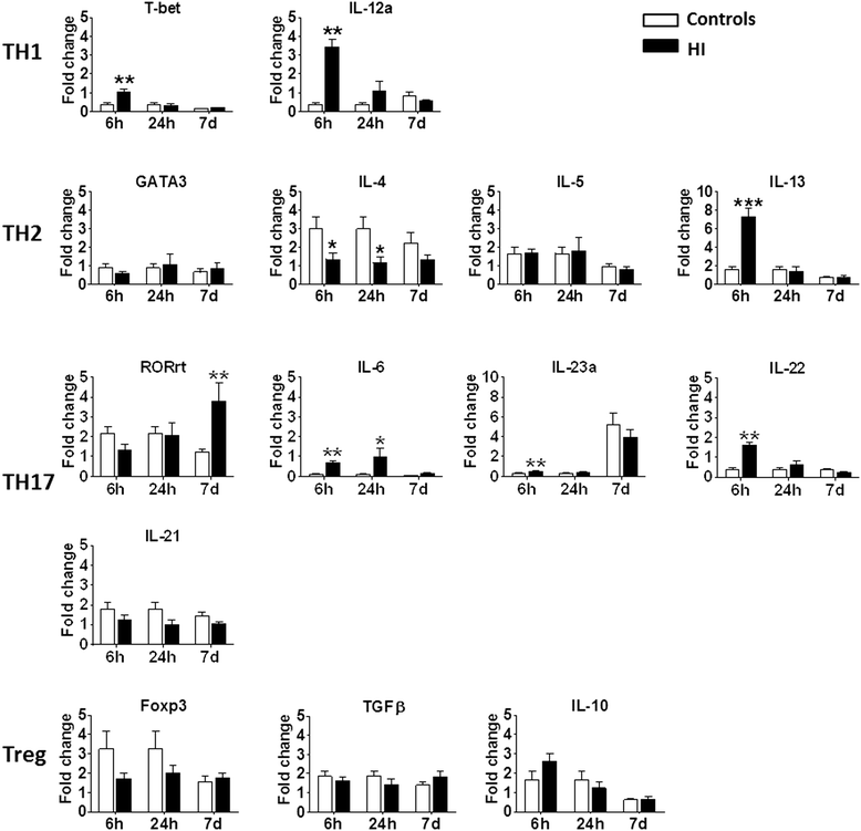
The canonical types of cytokine and transcription factor gene expression in the brain after hypoxia-ischemia (HI). *P < 0.05, **P < 0.01, ***P < 0.001 using Student’s unpaired t-test. Data are presented as mean ± SEM. N = 5 to 8/group. White bars: undamaged controls; Black bars: post-HI ipsilateral hemisphere. Abbreviations: Th: T-helper cells; Treg: T-regulatory cells.

References

    1. Pierson CR, Folkerth RD, Billiards SS, Trachtenberg FL, Drinkwater ME, Volpe JJ, Kinney HC. Gray matter injury associated with periventricular leukomalacia in the premature infant. Acta Neuropathol. 2007;114:619–631. doi: 10.1007/s00401-007-0295-5. - DOI - PMC - PubMed
    1. Leviton A, Gressens P. Neuronal damage accompanies perinatal white-matter damage. Trends Neurosci. 2007;30:473–478. doi: 10.1016/j.tins.2007.05.009. - DOI - PubMed
    1. Inder TE, Huppi PS, Warfield S, Kikinis R, Zientara GP, Barnes PD, Jolesz F, Volpe JJ. Periventricular white matter injury in the premature infant is followed by reduced cerebral cortical gray matter volume at term. Ann Neurol. 1999;46:755–760. doi: 10.1002/1531-8249(199911)46:5<755::AID-ANA11>3.0.CO;2-0. - DOI - PubMed
    1. Back SA, Miller SP. Brain injury in premature neonates: a primary cerebral dysmaturation disorder? Ann Neurol. 2014;75:469–486. doi: 10.1002/ana.24132. - DOI - PMC - PubMed
    1. Fleiss B, Gressens P. Tertiary mechanisms of brain damage: a new hope for treatment of cerebral palsy? Lancet Neurol. 2012;11:556–566. doi: 10.1016/S1474-4422(12)70058-3. - DOI - PubMed

Publication types