. 2015 Jan;33(1):133-45.
doi: 10.1002/stem.1841.

Glucose transporter 1-positive endothelial cells in infantile hemangioma exhibit features of facultative stem cells

Affiliations

Glucose transporter 1-positive endothelial cells in infantile hemangioma exhibit features of facultative stem cells

Lan Huang et al. Stem Cells. 2015 Jan.

Abstract

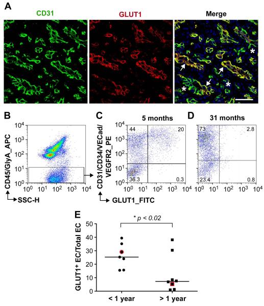
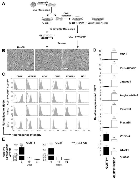
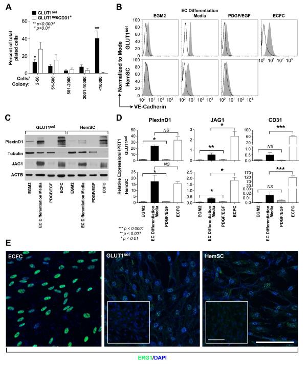
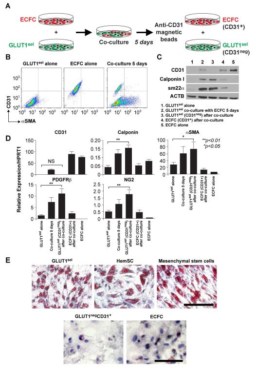
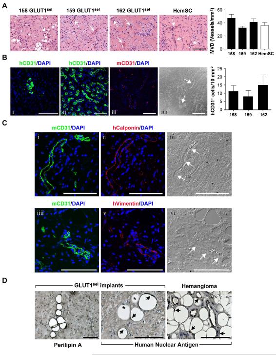
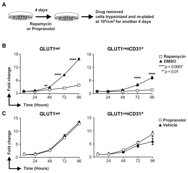
Endothelial glucose transporter 1 (GLUT1) is a definitive and diagnostic marker for infantile hemangioma (IH), a vascular tumor of infancy. To date, GLUT1-positive endothelial cells in IH have not been quantified nor directly isolated and studied. We isolated GLUT1-positive and GLUT1-negative endothelial cells from IH specimens and characterized their proliferation, differentiation, and response to propranolol, a first-line therapy for IH, and to rapamycin, an mTOR pathway inhibitor used to treat an increasingly wide array of proliferative disorders. Although freshly isolated GLUT1-positive cells, selected using anti-GLUT1 magnetic beads, expressed endothelial markers CD31, VE-Cadherin, and vascular endothelial growth factor receptor 2, they converted to a mesenchymal phenotype after 3 weeks in culture. In contrast, GLUT1-negative endothelial cells exhibited a stable endothelial phenotype in vitro. GLUT1-selected cells were clonogenic when plated as single cells and could be induced to redifferentiate into endothelial cells, or into pericytes/smooth muscle cells or into adipocytes, indicating a stem cell-like phenotype. These data demonstrate that, although they appear and function in the tumor as bona fide endothelial cells, the GLUT1-positive endothelial cells display properties of facultative stem cells. Pretreatment with rapamycin for 4 days significantly slowed proliferation of GLUT1-selected cells, whereas propranolol pretreatment had no effect. These results reveal for the first time the facultative nature of GLUT1-positive endothelial cells in IH.

Keywords: Endothelial cells; Facultative stem cells; Glucose transporter 1; Infantile hemangioma.


Figures

Figure 1
Figure 1. GLUT1+ and GLUT1neg endothelial cells in IH
(A): Anti-human CD31 (green) and anti-GLUT1 (red) staining of proliferating IH tissue section (arrows, GLUT1+ vessels; *, GLUT1neg vessels). Nuclei counterstained with DAPI (blue). Scale bar, 50 μm. Flow cytometric analyses of freshly isolated cells from proliferating IH ( 5 months old ) (B-C) and involuting IH (31 months old) (D) using antibodies against human CD45/GlyA (APC), CD31/CD34/VECad/VEGFR2 (PE) and GLUT1 (FITC). Percentages of cells in each quadrant are shown (C,D). (E) Percent GLUT1+ ECs, quantified by flow cytometry, in IH specimens (N=15). •, IH specimens from children under one year of age; ■, IH specimens from children over one year of age. The specimens shown in C and D are highlighted in red. * p <0.02 by unpaired t test.
Figure 2
Figure 2. GLUT1sel cells versus GLUT1negCD31+ cells
(A): Schematic of cell isolation using sequential antibody-coated magnetic beads. (B): Phase contrast image of GLUT1sel cells and GLUT1negCD31+ cells after 3 weeks in culture. HemSC are shown for comparison on the left. Scale bar, 100 μm. (C): Flow cytometric analysis of GLUT1sel cells and GLUT1negCD31+ cells for endothelial (CD31, VEGFR2), hematopoietic (CD45), and mesenchymal (CD90, PDGFRβ and NG2) markers. HemSC and ECFC included as controls. Black lines depict cells incubated with isotype-matched control antibodies; shaded grey areas are cells incubated with primary antibodies. (D): QPCR measured mRNA levels in GLUT1sel cells (black bars) and GLUT1negCD31+ cells (white bars) after 3 weeks in culture (N=3). (E): QPCR measured mRNA levels of GLUT1 and CD31 in freshly anti-GLUT1 magnetic bead selected GLUT1+ cells on day0 and GLUT1sel cells on day10 and 24 in the culture. * p <0.01 and *** p <0.0001 by paired t test.
Figure 3
Figure 3. GLUT1sel cells are clonogenic and can be induced to undergo endothelial differentiation in vitro
(A): GLUT1sel cells (black bars) form larger colonies than GLUT1negCD31+ cells (white bars) in single cell clonogenic assays (N = 5). *P<0.01, **P<0.0001 by paired t test. (B-C): GLUT1sel cells and HemSC induced to re-express endothelial markers after 5 days in EC differentiation media (N=4). Endothelial markers VE-Cadherin examined by flow cytometry (B) and PlexinD1 and Jagged1 (JAG1) examined by western blot (C). In B, black lines indicate cells incubated with isotype-matched control antibodies; shaded grey area, cells incubated with anti-VE-Cadherin antibody. ECFCs served as endothelial positive control in both. Cells cultured for 5 days in EGM-2 and in EC differentiation media with PDGF-BB and EGF served as negative controls. Tubulin or ACTB served as the loading control for the western blot. (D): QPCR for PlexinD1, Jagged1 (JAG1) and CD31 in GLUT1sel cells and HemSC after 5 days of EC differentiation. ECFCs served as endothelial positive control. Cells cultured for 5 days in EGM-2 and in EC differentiation media with PDGFBB and EGF were negative controls. *P<0.01, **P<0.001 and ***P<0.0001 by ANOVA and unpaired t test. (E): Immunofluorescence staining for ERG1 in GLUT1sel cells and HemSC after 5 days of EC differentiation. ECFC served as endothelial positive control. Insets are GLUT1sel cells and HemSC that were not stimulated to undergo EC differentiation. Scale bar, 50 μm.
Figure 4
Figure 4. GLUT1sel cells can be induced to express pericytic/SMC phenotype and undergo adipogenesis
(A): Schematic of pericyte/SMC differentiation assay: 5 days of co-culture with ECFCs, followed by removal with anti-CD31-coated magnetic beads. (B): CD31 and αSMA expression measured by flow cytometry of saponin-permeabilized cells. (C): Calpoinin I and sm22α measured by western blot. CD31 shown to evaluate ECFC removal. ACTB used as the loading control. (D): QPCR to analyze mRNA levels of pericyte/SMC markers. N=4, *P<0.05 and **P<0.01 by ANOVA followed by paired t test. (E): Cells induced to undergo adipogenesis stained with Oil Red-O. HemSC and mesenchymal stem cells served as positive controls; ECFC served as negative control. Scale bar, 80 μm. Results are representative of independent assays using paired GLUT1sel and GLUT1negCD31+ cells isolated from 4 different IH specimens.
Figure 5
Figure 5. GLUT1sel cells form ECs, pericytes/SMCs and adipocytes in immunodeficient mice
GLUT1sel cells from 3 different IH (158,159 and 162) mixed with Matrigel and injected subcutaneously into nu/nu mice for 14 days. (A): H&E staining shows microvessels in the implants (white arrows indicate lumens with red blood cells). HemSC are shown for comparison. Scar bar, 150 μm. Microvessel density (MVD) quantified (N=5) and compared to HemSC [10]. (B): anti-human CD31 (green, i) staining of GLUT1sel cell/Matrigel implants compared to anti-human CD31 staining of human IH specimen (green, ii). GLUT1sel cell/Matrigel implant stained with anti-mouse CD31 (red, iii) with corresponding phase contrast image (iiii). White arrow indicates blood vessel lumen. Scale bar, 100 μm. Human CD31+ cells in the implants were quantified (N=5) (C): Serial sections from GLUT1sel cell/Matrigel implant stained with anti-mouse CD31 (green, i) and anti-human Calponin (red, ii) with corresponding phase contrast image (iii). Serial sections from these implants stained with anti-mouse CD31 (green, iiii) and anti-human Vimentin (red, v), with corresponding phase contrast image (vi). White arrows indicate blood vessel lumen. Scale bar, 100 μm. (D): Anti-perilipin A staining of GLUT1sel cell/Matrigel implants (left) panel and anti-human nuclear antigen staining of GLUT1sel /Matrigel implants and involuting IH specimen (middle and right panels). Arrows indicate adipocytes stained with anti-human nuclei; * indicate non-stained adipocytes. Scale bar, 100 μm.
Figure 6
Figure 6. Rapamycin suppresses proliferation of GLUT1sel cells and GLUT1negCD31+ cells in vitro
(A): Schematic of the experiment. (B): Proliferation of GLUT1sel and GLUTnegCD31+ cells in “drug-free” culture after 4-day pretreatment with rapamycin (B) or propranolol (C). Cells pretreated with DMSO (▲) in Panel B or vehicle (▲) in Panel C served as controls. N=3, **P<0.01 and ****P<0.0001 by ANOVA.

References

    1. Enjolras O, Mulliken JB. The current management of vascular birthmarks. Pediatr Dermatol. 1993;10:311–313. - PubMed
    1. Frieden IJ, Haggstrom AN, Drolet BA, et al. Infantile hemangiomas: current knowledge, future directions. Proceedings of a research workshop on infantile hemangiomas, April 7-9, 2005, Bethesda, Maryland, USA. Pediatr Dermatol. 2005;22:383–406. - PubMed
    1. North PE, Waner M, Mizeracki A, et al. GLUT1: a newly discovered immunohistochemical marker for juvenile hemangiomas. Hum Pathol. 2000;31:11–22. - PubMed
    1. North PE, Waner M, Mizeracki A, et al. A unique microvascular phenotype shared by juvenile hemangiomas and human placenta. Arch Dermatol. 2001;137:559–570. - PubMed
    1. Boye E, Yu Y, Paranya G, et al. Clonality and altered behavior of endothelial cells from hemangiomas. J Clin Invest. 2001;107:745–752. - PMC - PubMed

Publication types

MeSH terms