. 2014 Oct 17;289(42):29285-96.
doi: 10.1074/jbc.M114.563924. Epub 2014 Sep 3.

Translocation of glycogen synthase kinase-3β (GSK-3β), a trigger of permeability transition, is kinase activity-dependent and mediated by interaction with voltage-dependent anion channel 2 (VDAC2)

Affiliations

Translocation of glycogen synthase kinase-3β (GSK-3β), a trigger of permeability transition, is kinase activity-dependent and mediated by interaction with voltage-dependent anion channel 2 (VDAC2)

Masaya Tanno et al. J Biol Chem. .

Abstract

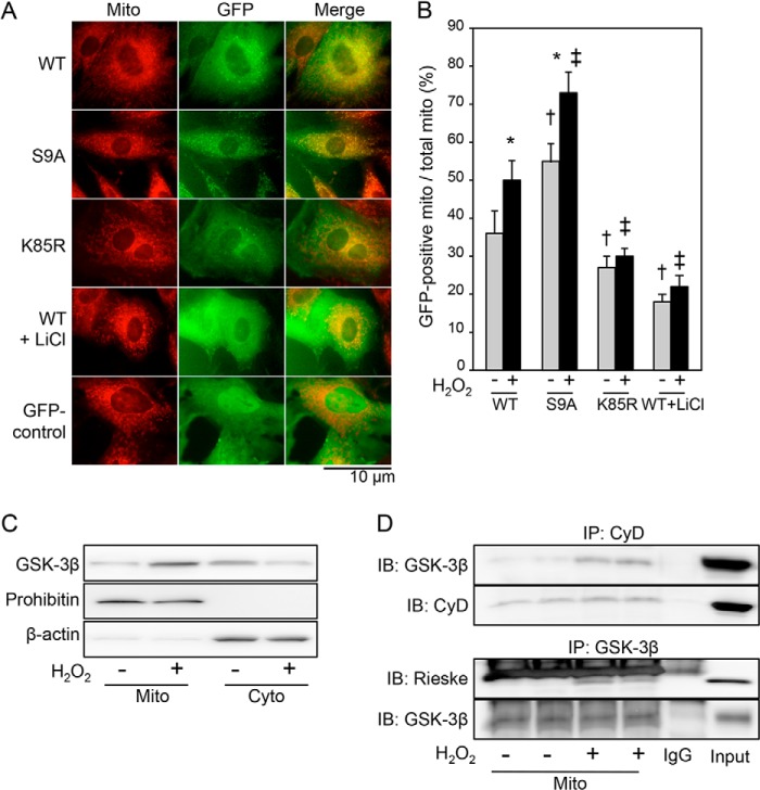
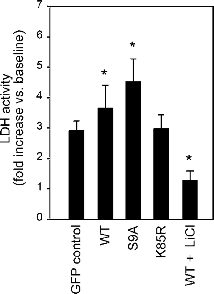
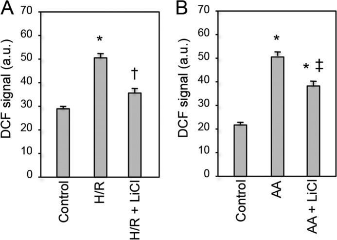
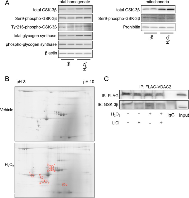
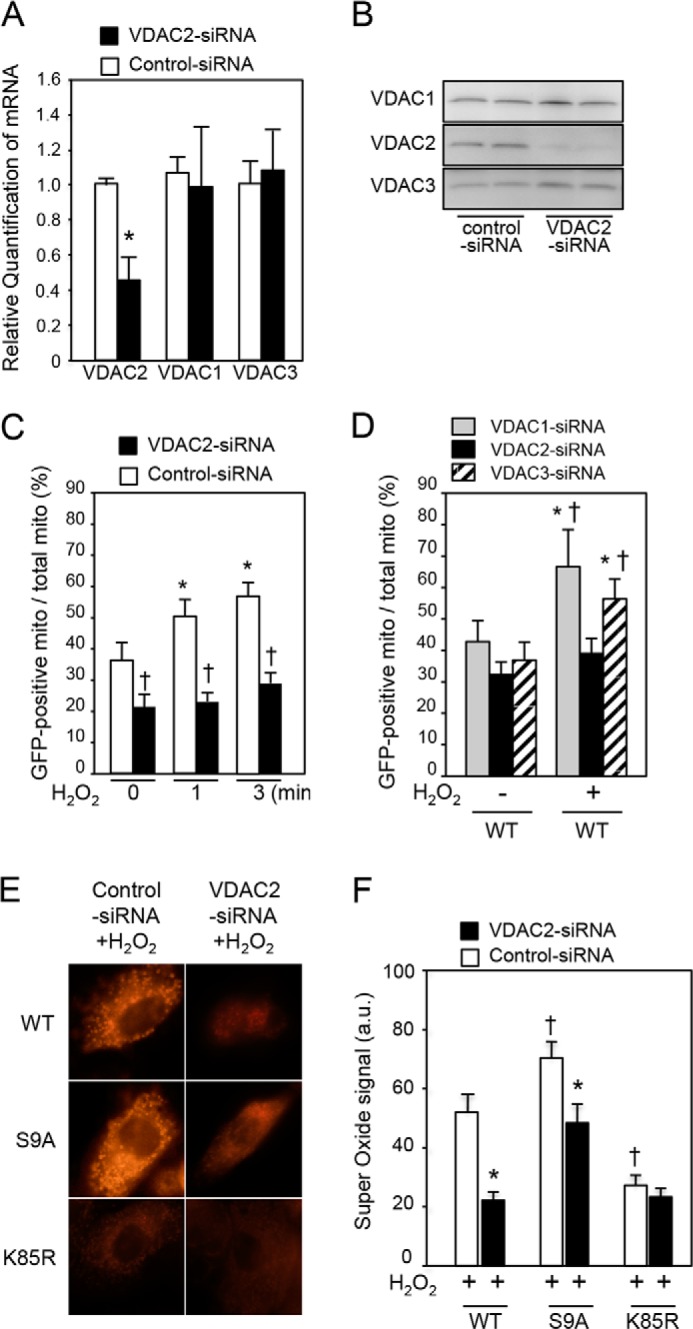
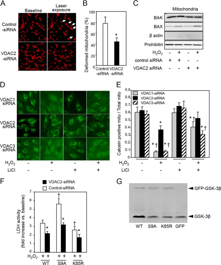
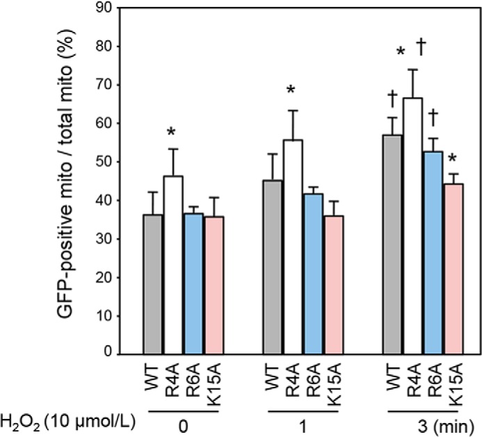
Glycogen synthase kinase-3β (GSK-3β) is a major positive regulator of the mitochondrial permeability transition pore (mPTP), a principle trigger of cell death, under the condition of oxidative stress. However, the mechanism by which cytosolic GSK-3β translocates to mitochondria, promoting mPTP opening, remains unclear. Here we addressed this issue by analyses of the effect of site-directed mutations in GSK-3β on mitochondrial translocation and protein/protein interactions upon oxidative stress. H9c2 cardiomyoblasts were transfected with GFP-tagged GSK-3β (WT), a mutant GSK-3β insensitive to inhibitory phosphorylation (S9A), or kinase-deficient GSK-3β (K85R). Time lapse observation revealed that WT and S9A translocated from the cytosol to the mitochondria more promptly than did K85R after exposure to oxidative stress. H2O2 increased the density of nine spots on two-dimensional gel electrophoresis of anti-GSK-3β-immunoprecipitates by more than 3-fold. MALDI-TOF/MS analysis revealed that one of the spots contained voltage-dependent anion channel 2 (VDAC2). Knockdown of VDAC2, but not VDAC1 or VDAC3, by siRNA attenuated both the mitochondrial translocation of GSK-3β and mPTP opening under stress conditions. The mitochondrial translocation of GSK-3β was attenuated also when Lys-15, but not Arg-4 or Arg-6, in the N-terminal domain of GSK-3β was replaced with alanine. The oxidative stress-induced mitochondrial translocation of GSK-3β was associated with an increase in cell death, which was suppressed by lithium chloride (LiCl), a GSK-3β inhibitor. These results demonstrate that GSK-3β translocates from the cytosol to mitochondria in a kinase activity- and VDAC2-dependent manner in which an N-terminal domain of GSK-3β may function as a mitochondrial targeting sequence.

Keywords: Glycogen Synthase Kinase 3 (GSK-3); Mitochondrial Permeability Transition (MPT); Mitochondrial Transport; Necrosis (Necrotic Death); Protein-Protein Interaction; Voltage-dependent Anion Channel 2.


Figures

FIGURE 1.
FIGURE 1.
Kinase activity-dependent mitochondrial translocation of GSK-3β under the condition of oxidative stress. A, fluorescence images obtained from time lapse observation. H9c2 cells were transfected with GFP-tagged GSK-3β (WT), constitutively active mutant GSK-3β (S9A), kinase-inactive mutant GSK-3β (K85R), or GFP alone (GFP control) and then stained with MitoTracker Red. Photos at 3 min after exposure of cells to H2O2 (10 μmol/liter) in the presence or absence of LiCl (30 mmol/liter) are shown. B, quantification of mitochondrial (mito) localization of each plasmid. MitoTracker-stained area overlapped with GFP signal was expressed as a percentage of total MitoTracker-stained area. *, p < 0.05 versus cells transfected with the same plasmid and treated with a vehicle; †, p < 0.05 versus cells transfected with WT and treated with a vehicle; ‡, p < 0.05 versus cells transfected with WT and exposed to H2O2. C, representative immunoblotting for total GSK-3β in mitochondrial fractions (Mito) and cytosolic fractions (Cyto). Prohibitin and β-actin were used as markers of the mitochondria and cytosol, respectively. Three separate experiments showed similar results. D, interaction of GSK-3β with mitochondrial proteins. Immunoblots (IB) for GSK-3β co-immunoprecipitated (IP) with cyclophilin D (CyD) and Rieske co-immunoprecipitated with GSK-3β with or without exposure to H2O2 (100 μmol/liter; 4 h) are shown. Error bars represent S.E.
FIGURE 2.
FIGURE 2.
Activity of GSK-3β is associated with cell death. LDH activity in the culture medium was measured at 4 h after addition of H2O2 (100 μmol/liter). -Fold increases in LDH activity compared with the baseline value are shown. *, p < 0.05 versus GFP control. Error bars represent S.E.
FIGURE 3.
FIGURE 3.
Effects of GSK-3β inhibition on ROS production by oxidative stress. Production of ROS after hypoxia/reoxygenation (A) or exposure to 50 μmol/liter antimycin A (B) was monitored by DCF staining. GSK-3β was inhibited by lithium chloride. Signal intensities of DCF staining are shown. *, p < 0.05 versus control; †, p < 0.05 versus hypoxia/reoxygenation (H/R); ‡, p < 0.05 versus antimycin A (AA). Error bars represent S.E. a.u., arbitrary units.
FIGURE 4.
FIGURE 4.
Effects of H2O2 on GSK-3β activity and its interaction with other proteins. A, H9c2 cells were exposed to 100 μmol/liter H2O2 or a vehicle (Ve) for 4 h and harvested. Results of Western blotting for total GSK-3β, Ser-9-phospho-GSK-3β, Tyr-216-phospho-GSK-3β, total glycogen synthase, and phospho-glycogen synthase in the total homogenate (left panels) and total GSK-3β and Ser-9-phospho-GSK-3β in the mitochondrial fraction (right panels) are shown. β-Actin and prohibitin serve as loading controls. B, representative two-dimensional gels stained by Coomassie Blue. Samples were obtained from GSK-3β immunoprecipitates of H9c2 cells that were treated with a vehicle or H202 (100 μmol/liter) for 3 h. Results of four experiments for vehicle-treated cells and four experiments for H2O2-exposed cells were similar. Spots that exhibited increases in the density by 3-fold or more after exposure to H2O2 are labeled as 1–9. C, HEK293 cells transfected with FLAG-VDAC2 were exposed to H2O2 (100 μmol/liter; 4 h) in the presence or absence of LiCl (30 mmol/liter). Immunoblots (IB) for FLAG and GSK-3β co-immunoprecipitated (IP) with FLAG-VDAC2 are shown.
FIGURE 5.
FIGURE 5.
VDAC2-dependent mitochondrial translocation of GSK-3β and mitochondrial production of superoxide. A and B, mRNA levels in H9c2 cells (A) and protein levels in HEK293 cells (B) of VDAC1, VDAC2, and VDAC3 at 48 h after transfection of VDAC2 siRNA. *, p < 0.05 versus control siRNA. C, mitochondrial localization of GFP-tagged GSK-3β (WT) expressed as a percentage of GFP-positive mitochondria among total mitochondria at baseline and at 1 and 3 min after exposure to H2O2 in H9c2 cells. *, p < 0.05 versus baseline; †, p < 0.05 versus control siRNA. D, HEK293 cells were transfected with VDAC1 siRNA, VDAC2 siRNA, or VDAC3 siRNA. Mitochondrial localization of GFP-tagged GSK-3β (WT) expressed as a percentage of GFP-positive mitochondria (mito) among total mitochondria with or without exposure to H2O2 (10 μmol/liter; 3 min) is shown. *, p < 0.05 versus baseline; †, p < 0.05 versus VDAC2 siRNA. E, effects of VDAC2 and kinase activity of GSK-3β on generation of superoxide. Superoxide signal after exposure to H2O2 in H9c2 cells transfected with WT, S9A, or K85R is shown. F, quantitative analysis of superoxide-positive pixels per cell is shown. *, p < 0.05 versus control siRNA; †, p < 0.05 versus WT. Error bars represent S.E. a.u., arbitrary units.
FIGURE 6.
FIGURE 6.
Effects of VDAC2 knockdown on mitochondrial deformation, mPTP opening, and cell necrosis by oxidant stress. A, photos of mitochondria obtained from time lapse observation by super-resolution microscopy (N-SIM). H9c2 cells were transfected with control siRNA or VDAC2 siRNA and then stained with MitoTracker Red before observation by N-SIM. Arrows indicate swollen mitochondria after intermittent laser scanning for time lapse observation. B, percentage of swollen or fragmented mitochondria after 4-min observation by N-SIM. *, p < 0.05 versus control siRNA. C, immunoblots for BAK and BAX in the mitochondrial fraction are shown. Prohibitin served as a loading control. Successful mitochondrial fractionation is indicated by clear prohibitin bands and barely detectable β-actin bands. D, open-closed status of mPTPs determined by calcein assay in HEK293 cells. Images of calcein-stained cells transfected with VDAC1 siRNA, VDAC2 siRNA, or VDAC3 siRNA in the presence or absence of H2O2 and/or LiCl are shown. E, the ratio of calcein-positive area to MitoTracker-positive area is shown as an index for mitochondria (mito) that were not subjected to opening of mPTPs. *, p < 0.05 versus baseline (without H2O2 and LiCl); †, p < 0.05 versus VDAC2 siRNA. F, LDH activity in the culture medium was measured at 4 h after addition of H2O2 (100 μmol/liter). -Fold increases in LDH activity compared with the baseline value are shown. G, Western blotting for GSK-3β in H9c2 cells transfected with GFP-GSK-3β construct (WT, S9A, or K85R) or GFP control. Endogenous GSK-3β and GFP-tagged GSK-3β constructs were detected around 46 and 79 kDa, respectively. Error bars represent S.E.
FIGURE 7.
FIGURE 7.
Effects of mutations in the N-terminal domain of GSK-3β on mitochondrial translocation. Percentages of GFP-positive mitochondria (mito) among total mitochondria at baseline and at 1 and 3 min after H2O2 exposure are shown. *, p < 0.05 versus WT; †, p < 0.05 versus baseline. Error bars represent S.E.

References

    1. Miura T., Tanno M. (2012) The mPTP and its regulatory proteins: final common targets of signalling pathways for protection against necrosis. Cardiovasc. Res. 94, 181–189 - PubMed
    1. Weiss J. N., Korge P., Honda H. M., Ping P. (2003) Role of the mitochondrial permeability transition in myocardial disease. Circ. Res. 93, 292–301 - PubMed
    1. Halestrap A. P. (2009) What is the mitochondrial permeability transition pore? J Mol. Cell. Cardiol. 46, 821–831 - PubMed
    1. Miura T., Tanno M., Sato T. (2010) Mitochondrial kinase signalling pathways in myocardial protection from ischaemia/reperfusion-induced necrosis. Cardiovasc. Res. 88, 7–15 - PubMed
    1. Halestrap A. P. (2010) A pore way to die: the role of mitochondria in reperfusion injury and cardioprotection. Biochem. Soc. Trans. 38, 841–860 - PubMed

Publication types

MeSH terms

Substances

LinkOut - more resources