. 2014 Nov;88(22):13135-48.
doi: 10.1128/JVI.01883-14. Epub 2014 Sep 3.

Induction of viral, 7-methyl-guanosine cap-independent translation and oncolysis by mitogen-activated protein kinase-interacting kinase-mediated effects on the serine/arginine-rich protein kinase

Affiliations

Induction of viral, 7-methyl-guanosine cap-independent translation and oncolysis by mitogen-activated protein kinase-interacting kinase-mediated effects on the serine/arginine-rich protein kinase

Michael C Brown et al. J Virol. 2014 Nov.

Abstract

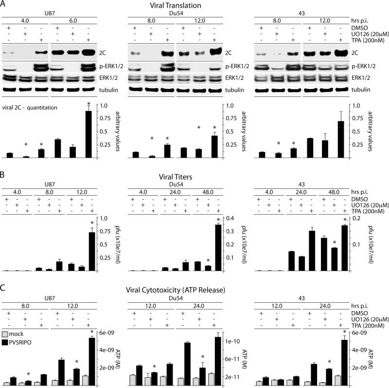
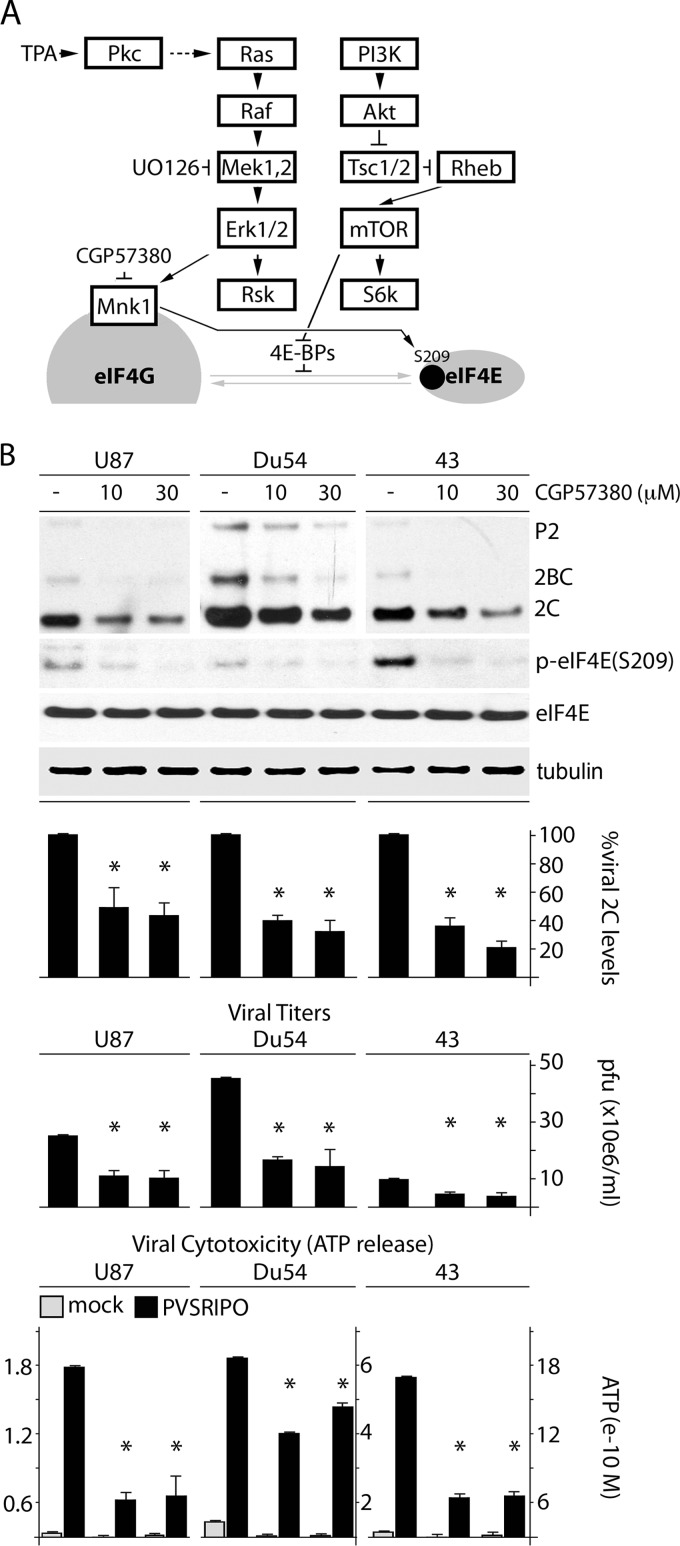
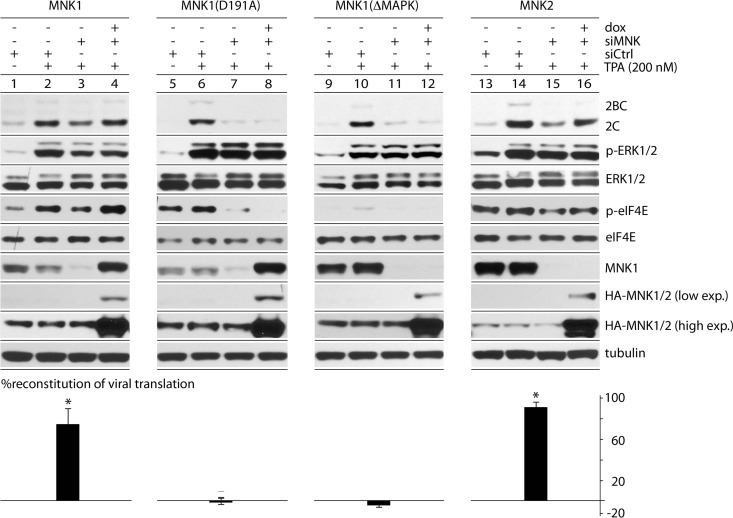
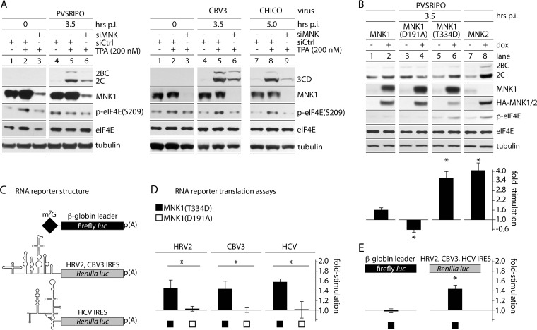
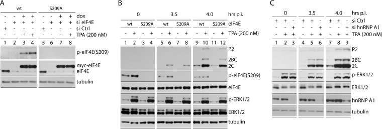
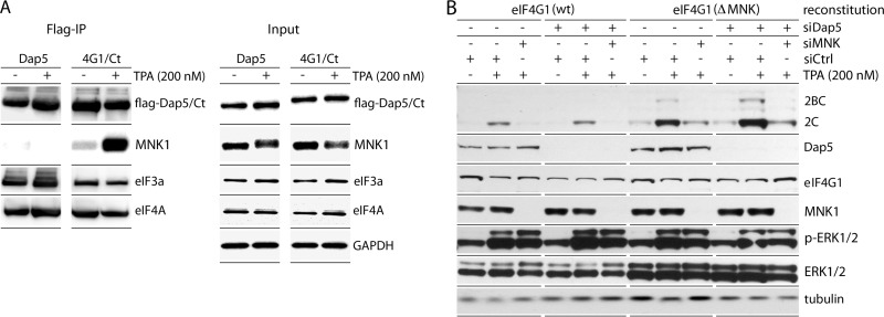
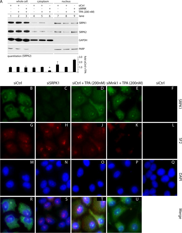
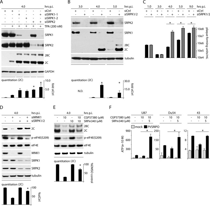
Protein synthesis, the most energy-consuming process in cells, responds to changing physiologic priorities, e.g., upon mitogen- or stress-induced adaptations signaled through the mitogen-activated protein kinases (MAPKs). The prevailing status of protein synthesis machinery is a viral pathogenesis factor, particularly for plus-strand RNA viruses, where immediate translation of incoming viral RNAs shapes host-virus interactions. In this study, we unraveled signaling pathways centered on the ERK1/2 and p38α MAPK-interacting kinases MNK1/2 and their role in controlling 7-methyl-guanosine (m(7)G) "cap"-independent translation at enterovirus type 1 internal ribosomal entry sites (IRESs). Activation of Raf-MEK-ERK1/2 signals induced viral IRES-mediated translation in a manner dependent on MNK1/2. This effect was not due to MNK's known functions as eukaryotic initiation factor (eIF) 4G binding partner or eIF4E(S209) kinase. Rather, MNK catalytic activity enabled viral IRES-mediated translation/host cell cytotoxicity through negative regulation of the Ser/Arg (SR)-rich protein kinase (SRPK). Our investigations suggest that SRPK activity is a major determinant of type 1 IRES competency, host cell cytotoxicity, and viral proliferation in infected cells.

Importance: We are targeting unfettered enterovirus IRES activity in cancer with PVSRIPO, the type 1 live-attenuated poliovirus (PV) (Sabin) vaccine containing a human rhinovirus type 2 (HRV2) IRES. A phase I clinical trial of PVSRIPO with intratumoral inoculation in patients with recurrent glioblastoma (GBM) is showing early promise. Viral translation proficiency in infected GBM cells is a core requirement for the antineoplastic efficacy of PVSRIPO. Therefore, it is critically important to understand the mechanisms controlling viral cap-independent translation in infected host cells.


Figures

FIG 1
FIG 1
ERK1/2 signals control PVSRIPO translation, proliferation, and cytotoxicity in GBM cells. (A) U87, Du54, or 43 GBM cells were treated with DMSO (mock), UO126 (20 μM), or TPA (200 nM) (30 min); infected with PVSRIPO; and harvested at the designated intervals. UO126 was maintained after infection; TPA was not. The immunoblots track viral protein (2C); the quantitation represents the average of 3 experiments normalized to the first control value for each series. (B) Supernatants from cells treated and infected as described for panel A were collected to determine viral progeny (PFU); the averages of two experiments are shown. (C) ATP release was measured in supernatants from the experiments in panel B. The ATP concentration was determined using a standard curve; the average of two assays is shown. The error bars represent SEM; the asterisks indicate significant ANOVA-protected t tests.
FIG 2
FIG 2
MNK inhibition reduces PVSRIPO translation, proliferation, and cytotoxicity in GBM cells. (A) Schema of signaling pathways to translation initiation factors investigated in this study. (B) U87, Du54, and 43 GBM cell lines were pretreated (1 h) with DMSO or CGP57380 (10 or 30 μM), followed by infection in the presence of DMSO/CGP57380. Cells were harvested (6 h p.i.), and viral protein (2C) was assessed by immunoblotting. Supernatants were collected (12 h p.i.) to determine viral progeny and ATP release, as indicated. The bars represent the average of 3 experiments normalized to the control (DMSO) values, and the error bars represent SEM. The asterisks denote significant ANOVA-protected t tests.
FIG 3
FIG 3
TPA-mediated stimulation of viral translation depends on catalytically active MNK. Dox-inducible cells expressing wt HA-tagged MNK1, MNK1(D191A), MNK1(ΔMAPK), or MNK2 (see the text for details) were used for mock or TPA stimulation after siCtrl (mock) or siMNK (MNK1) depletion. Dox-induced expression (exp.) of the corresponding HA-tagged MNK (lanes 4, 8, 12, and 16) was detected by HA immunoblotting. The MNK1 antibody used does not recognize MNK1(ΔMAPK) or MNK2. Viral translation was assayed at 4 h p.i. The percent reconstitution of viral translation was calculated as follows: (siMNK + Dox + TPA − siMNK + TPA) divided by (siCtrl + TPA − siMNK + TPA), representing the average of 3 experiments. The error bars represent SEM, and the asterisks indicate significance (P < 0.05 by Student's t test compared to 0% reconstitution).
FIG 4
FIG 4
MNK activity selectively enhances viral IRES-mediated translation. (A) HeLa cells were treated with control (siCtrl) or MNK1-targeting (siMNK) siRNA 24 h prior to TPA/mock stimulation. PVSRIPO (left) or CBV3/CHICO (right) infection and subsequent analyses were carried out as for Fig. 1. The assays were repeated 3 times, and representative series are shown. (B) Cells with Dox-inducible expression of wt MNK1, MNK1(D191A), MNK1(T334D), or MNK2 were mock/Dox induced (12 h) and infected with PVSRIPO. Viral translation was quantitated, and the fold stimulation of viral translation was calculated by dividing the Dox-induced value by the mock-induced value for 3 independent tests. (C) Structure of RNA reporters used (32). (D) Uncapped, in vitro-transcribed rluc RNA reporters driven by the HRV2, CBV3, or HCV 5′ UTR were cotransfected with m7G-capped β-globin leader fluc reporters into Dox-/mock-induced MNK1(T334D)- or MNK1(D191A)-expressing cells (4 h). IRES-driven (rluc) values were divided by β-globin 5′-UTR firefly luciferase values to correct for transfection differences. Dox-induced values were then divided by mock-induced values for each cell line to calculate the fold stimulation of IRES-mediated translation due to MNK1(T334D)/MNK1(D191A) expression. The data represent 3 independent assays done in triplicate for each cell line and 5′ UTR. (E) Pooled β-globin 5′-UTR fluc values and IRES-driven rluc values in MNK1(T334D)-expressing cells. (B, D, and E) The error bars represent SEM, and the asterisks indicate significance (P < 0.05 by Student's t test).
FIG 5
FIG 5
eIF4E(S209A) substitution and hnRNP A1 depletion do not affect TPA stimulation of PVSRIPO translation. (A) Dox-inducible cell lines expressing wt myc-eIF4E or myc-eIF4E(S209A) were treated with siCtrl (lanes 1 and 5) or eIF4E-targeting siRNA (lanes 2 to 4 and 6 to 8). Dox induction reconstituted wt eIF4E or eIF4E(S209A) to roughly endogenous levels. (B) The assay conditions from panel A, lanes 3 and 4 and lanes 7 and 8, were used to track viral translation after TPA stimulation in the presence of wt myc-eIF4E or myc-eIF4E(S209A) at 3.5 and 4 h p.i. (C) HeLa cells were mock or TPA stimulated following siCtrl or hnRNP A1 siRNA, as shown. The cells were infected, and viral protein was analyzed by immunoblotting (3.5 and 4 h p.i.). The experiments were performed in triplicate (B) or duplicate (C); representative series are shown.
FIG 6
FIG 6
MNK1 stimulation of viral translation does not require MNK1 binding to eIF4G1. (A) Dox-inducible endogenous eIF4G1 knockdown with simultaneous mock reconstitution (pcDNA5) or reconstitution with wt myc-eIF4G1-Flag or myc-eIF4G1(ΔMNK)-Flag. (B) Flag IP of lysates from the three cell lines after Dox induction (96 h). MNK1 co-IP occurred only with wt eIF4G1 reconstituted cells. (C) MNK1/mock depletion, TPA/mock stimulation, and PVSRIPO infection of the three cell lines as shown in Fig. 4A. Prior to infection, all cells were Dox induced (96 h), followed by siCtrl/siMNK1 treatment and TPA/mock stimulation, as shown. Note deficient eIF4E(S209) phosphorylation in mock-/eIF4G(ΔMNK)-reconstituted cells. Viral protein 2C levels were quantitated (4 h p.i.), and the values represent the average of 3 experiments normalized using the corresponding siCtrl-mock lanes. The error bars indicate SEM.
FIG 7
FIG 7
Dap5 does not interact with MNK and does not affect MNK-dependent PVSRIPO translation competence. (A) HEK293 cells with Dox-inducible expression of Dap5-Flag or eIF4G1(682-1600)-Flag (Ct) (8) were treated with Dox (24 h) and subsequently mock (DMSO) or TPA (200 nM) stimulated (2 h). Flag IP of Dap5/eIF4G1(682-1600) and co-IP of MNK1, eIF4A, and eIF3a are shown. The experiment was repeated 10 times; a representative series is shown. A shift in MNK1 electrophoretic mobility after TPA stimulation likely reflects phosphorylation. (B) HeLa (endogenous) eIF4G1 knockdown/(exogenous) wt eIF4G1 or eIF4G1(ΔMNK) knock-in cells were treated as for Fig. 5. In addition, the cells were treated with siCtrl or siDap5 (72 h) prior to infection. PVSRIPO translation was assayed for each condition alongside relevant controls. The assay was repeated twice, and a representative series is shown.
FIG 8
FIG 8
MNK1 depletion causes SRPK nuclear accumulation and SF2 nuclear-speckle dissociation. (A) Whole-cell, cytoplasmic, and nuclear lysates were prepared from cells treated with siCtrl with and without TPA (1 h) or siMNK plus TPA (1 h). Immunoblots for SRPK2 were quantitated, and average SRPK2 values for each fraction from two experiments are shown; the asterisk denotes an ANOVA-protected t test. The error bars indicate SEM. (B to U) Indirect IF using cells treated with siCtrl (with or without TPA; 1 h), siMNK (plus TPA; 1 h), or siSRPK1 were stained for SRPK1 (green), nuclear-speckle marker SF2 (red), and DAPI (blue), as labeled. Individual staining for each group (B to Q) and merged tricolor staining (R to U) are shown. A negative-staining (no primary antibody added) control for SRPK1 and SF2 is shown on the right (F, L, and Q). The indirect IF experiment was performed three times, and representative images are shown.
FIG 9
FIG 9
SRPK depletion enhances PVSRIPO translation and propagation, counters the MNK depletion/inhibition effect, and increases viral translation and cytotoxicity in GBM. (A) HeLa cells were treated with siCtrl (with or without TPA) or siRNA targeting SRPK1 and/or SRPK2 (all plus TPA) prior to infection with PVSRIPO (4 h). The quantitation represents the average viral protein 2C levels normalized for the siCtrl-plus-TPA sample from 2 assays; asterisks denote ANOVA-protected t test. (B) HeLa cells were treated with siCtrl or siRNA targeting SRPK1 and -2 and assessed for viral 2C expression by immunoblotting at 4 and 5 h p.i. Average quantitations from 3 tests, normalized for the control values for each interval, are shown. (C) Viral titers from HeLa cells treated as for panel B and infected at an MOI of 10 were determined for the designated intervals; the average of two experiments is shown, normalizing between experiments using the siCtrl values. (D) HeLa cells were cotransfected with siCtrl or siRNA targeting SRPK1/2 followed by transfection with siCtrl or MNK1 siRNA as shown and infected as for panel B. The quantitation represents the average viral protein 2C levels normalized between experiments by setting siCtrl values to 1. (E) HeLa cells were treated with DMSO (mock), CGP57380 (10 μM), SRPin340 (10 μM), or combined inhibitors coincident with PVSRIPO infection (4 h p.i.). Cells were harvested and analyzed for viral protein by immunoblotting. Quantitation of viral protein 2C is shown for 3 assays normalized by setting the controls (without CGP57380) to 1. (F) U87, Du54, and 43 GBM cells were treated with DMSO, CGP57380, or CGP57380 plus SRPin340 at the concentrations indicated at the time of infection. The supernatants were harvested (12 h p.i.), and the average ATP concentration for 3 (Du54) or 2 (U87 and 43) assays was determined. (B to F) The asterisks denote significant paired Student's t tests; error bars represent SEM.

References

    1. Kozak M. 1991. An analysis of vertebrate mRNA sequences: intimations of translational control. J. Cell Biol. 115:887–903. 10.1083/jcb.115.4.887. - DOI - PMC - PubMed
    1. Marintchev A, Edmonds KA, Marintcheva B, Hendrickson E, Oberer M, Suzuki C, Herdy B, Sonenberg N, Wagner G. 2009. Topology and regulation of the human eIF4A/4G/4H helicase complex in translation initiation. Cell 136:447–460. 10.1016/j.cell.2009.01.014. - DOI - PMC - PubMed
    1. Sweeney TR, Abaeva IS, Pestova TV, Hellen CU. 2014. The mechanism of translation initiation on type 1 picornavirus IRESs. EMBO J. 33:76–92. 10.1002/embj.201386124. - DOI - PMC - PubMed
    1. Pelletier J, Sonenberg N. 1988. Internal initiation of translation of eukaryotic mRNA directed by a sequence derived from poliovirus RNA. Nature 334:320–325. 10.1038/334320a0. - DOI - PubMed
    1. Jang SK, Krausslich HG, Nicklin MJ, Duke GM, Palmenberg AC, Wimmer E. 1988. A segment of the 5′ nontranslated region of encephalomyocarditis virus RNA directs internal entry of ribosomes during in vitro translation. J. Virol. 62:2636–2643. - PMC - PubMed

Publication types

MeSH terms

Substances