. 2014 Nov;88(22):13161-72.
doi: 10.1128/JVI.01766-14. Epub 2014 Sep 3.

Activation of NF-κB by the Kaposi's sarcoma-associated herpesvirus K15 protein involves recruitment of the NF-κB-inducing kinase, IκB kinases, and phosphorylation of p65

Affiliations

Activation of NF-κB by the Kaposi's sarcoma-associated herpesvirus K15 protein involves recruitment of the NF-κB-inducing kinase, IκB kinases, and phosphorylation of p65

Anika Hävemeier et al. J Virol. 2014 Nov.

Abstract

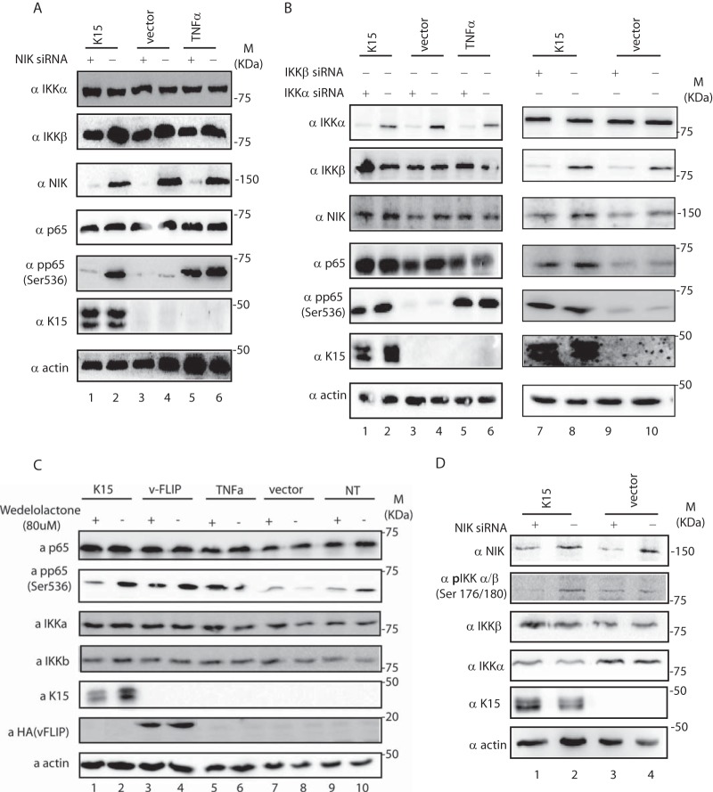
Kaposi's sarcoma herpesvirus (KSHV) (or human herpesvirus 8) is the cause of Kaposi's sarcoma, primary effusion lymphoma (PEL), and the plasma cell variant of multicentric Castleman's disease (MCD). The transmembrane K15 protein, encoded by KSHV, has been shown to activate NF-κB and the mitogen-activated protein kinases (MAPKs) c-jun-N-terminal kinase (JNK) and extracellular signal-regulated kinase (Erk) as well as phospholipase C gamma (PLCγ) and to contribute to KSHV-induced angiogenesis. Here we investigate how the K15 protein activates the NF-κB pathway. We show that activation of NF-κB involves the recruitment of NF-κB-inducing kinase (NIK) and IKK α/β to result in the phosphorylation of p65/RelA on Ser536. A K15 mutant devoid in NIK/IKK recruitment fails to activate NF-κB but remains proficient in the stimulation of both NFAT- and AP1-dependent promoters, showing that the structural integrity of the mutant K15 protein has not been altered dramatically. Direct recruitment of NIK represents a novel way for a viral protein to activate and manipulate the NF-κB pathway.

Importance: KSHV K15 is a viral protein involved in the activation of proinflammatory and angiogenic pathways. Previous studies reported that K15 can activate the NF-κB pathway. Here we show the molecular mechanism underlying the activation of this signaling pathway by K15, which involves direct recruitment of the NF-κB-inducing kinase NIK to K15 as well as NIK-mediated NF-κB p65 phosphorylation on Ser536. K15 is the first viral protein shown to activate NF-κB through direct recruitment of NIK. These results indicate a new mechanism whereby a viral protein can manipulate the NF-κB pathway.


Figures

FIG 1
FIG 1
K15-P induces the binding of NF-κB to target DNA sequence. (A) Left, schematic diagram of K15 and its functional domains. Right, alignment of the protein sequence of the K15-P and M cytoplasmic domains. The conserved motifs are underlined, and the motif in K15-P (aa 359 to 364), shown to be important in this report for the recruitment of NIK and IKKs, is shaded in gray. (B) HEK 293-T cells were cotransfected in duplicate with a vector control or increasing amounts (200 ng, 500 ng, 1 μg) of expression constructs for K15-P wt or K15-P Y481F and an NF-κB-responsive reporter vector. Forty hours after transfection, cells were lysed and the luciferase activity was measured. Equal expression levels of K15 were analyzed by immunoblotting. (C) Electrophoretic mobility shift assays were carried out with 32P-end-labeled double-stranded oligonucleotides corresponding to the consensus binding site for NF-κB (wt NF-κB) or with oligonucleotides containing a mutated NF-κB consensus binding site (mut NF-κB). Reactions were performed either without nuclear extracts (−) or with lysates from HeLa cells transfected with a vector control, K15-P wt, K15-P Y481F, NIK, or vFLIP expression constructs. The NF-κB-oligonucleotide complex is indicated as NF-κB-C. (D) Supershift analyses (SS) were performed with K15-P wt-transfected cells using antibodies specific for different NF-κB proteins (5 μg each), as well as 5 μg anti-IgG as an isotype control. Competition experiments were carried out with 200-fold molar excess of unlabeled wild-type (wt NF-κB) or mutated (mut NF-κB) oligonucleotides. All the experiments were performed three times. **, P < 0.01; ***, P < 0.001.
FIG 2
FIG 2
Silencing/inhibition of NIK and p65 inhibits K15-dependent NF-κB activation. (A and B) HEK 293 cells were transfected with the indicated siRNAs. Six hours later, the medium was replaced and cells were cotransfected with the indicated expression constructs and an NF-κB-responsive reporter vector. Forty hours after transfection, cells were lysed and the luciferase activity was measured. Shown are relative light units (RLUs) based on duplicate samples. Expression levels of different proteins were analyzed by immunoblotting. (C) HEK 293-T cells were transiently transfected with an NF-κB-responsive reporter vector, an empty vector, or a K15 expression vector and increasing amounts of dnNIK (500 ng and 1 μg). Forty hours after transfection, cells were lysed and luciferase activity was measured. Shown are relative light units based on duplicate samples. Expression levels of K15-P and dnNIK proteins were analyzed by immunoblotting. The experiment was performed three times in duplicate. **, P < 0.01; ***, P < 0.001.
FIG 3
FIG 3
K15 induces a NIK-dependent phosphorylation of p65 on Ser536. (A and B) HeLa cells were transfected with the indicated siRNAs. Six hours later, the medium was replaced and cells were transfected with the indicated expression constructs for K15, vFLIP, or vector control or left untransfected. Forty hours after transfection, cells were treated as indicated with TNF-α for 5 min and lysed in TBST buffer. Expression levels of different proteins were analyzed by immunoblotting. (C) HeLa cells were either transfected with 1 μg of the indicated expression construct or left untransfected, and 16 h later cells were treated with either wedelolactone or dimethyl sulfoxide (DMSO). Forty hours after transfection, cells were treated with TNF-α for 5 min as indicated and then lysed as described for panels A and B. Protein expression levels were analyzed by immunoblotting. (D) HeLa cells were treated as described for panel A and lysed in TBST buffer. Expression levels of the indicated proteins were measured by Western blotting. With the exception of the experiment shown in panel D, all other experiments were performed three times.
FIG 4
FIG 4
K15-P interacts directly with NIK, IKKα, and IKKβ via six amino acids near the last transmembrane domain of K15-P. (A) HEK 293-T cells were cotransfected with the indicated expression constructs or control vector and lysed 48 h later as described in Materials and Methods. Cleared cell lysates were used in a coimmunoprecipitation assay with either α-FLAG or mouse immunoglobulin protein G Sepharose beads. The beads were washed and analyzed by SDS-PAGE and Western blotting. (B) Schematic diagram of GST-K15 fusion proteins used in panels C to F. (C) GST fusion constructs, as depicted in panel B, were used in a GST pulldown assay with transiently expressed NIK or endogenous IKKα and IKKβ. GST proteins were incubated overnight with eukaryotic cell lysates. After extensive washing, beads were analyzed by SDS-PAGE and immunoblotting with an antibody against NIK, IKKα, or IKKβ. (D) NIK, IKKα, and IKKβ proteins were in vitro translated using the TNT coupled reticulocyte lysate system (Promega) and incubated overnight with K15-P fusion proteins or GST only, with which Sepharose beads were coated, at 4°C. The beads were washed, heated with sample buffer, and analyzed by SDS-PAGE and Western blotting. (E) K15-P alanine scanning mutants (see the text) in the background of GST-K15355–374 were used in a GST pulldown assay with transiently expressed NIK or endogenous IKKα and IKKβ. GST proteins were incubated overnight with eukaryotic cell lysates. After extensive washing, beads were analyzed by SDS-PAGE and immunoblotting with antibodies against NIK, IKKα, or IKKβ. (F) GST fusion proteins containing the entire K15 cytoplasmic domain for K15 wt and the indicated mutants were used in a GST pulldown assay with transiently expressed NIK or endogenous IKKα and IKKβ. GST proteins and eukaryotic cell lysates were incubated overnight. After extensive washing, beads were analyzed by SDS-PAGE and immunoblotting with antibodies against NIK, IKKα, or IKKβ. The experiments were performed three times.
FIG 5
FIG 5
K15 mutants deficient in binding to NIK, IKKα, and IKKβ show a decreased ability to activate NF-κB and induce p65 phosphorylation but retain the ability to activate other K15-P-driven promoters. (A) HEK 293-T cells were transiently cotransfected with reporter vectors responsive to NF-κB and increasing amounts (200 ng, 500 ng, 1 μg) of expression constructs of either K15-P wt, K15-P Y481F, K15-P 359–361 RQR/AAA, or K15-P362–364 RRR/AAA. Forty hours after transfection, cells were lysed and luciferase activity was measured. Shown are relative light units based on duplicate samples. Expression levels of K15-P wt and mutants were analyzed by immunoblotting. (B) Electrophoretic mobility shift assays were carried out with 32P-end-labeled double-stranded oligonucleotides corresponding to the consensus binding site for NF-κB. Reactions were performed either without nuclear extracts (−) or with lysates from HeLa cells transfected with empty vector, K15-P wt, K15-P359–361RQR/AAA, or K15-P362–364 RRR/AAA expression constructs. The NF-κB-oligonucleotide complex is indicated as NF-κB-C. (C) HeLa cells were transfected with the indicated expression constructs or left untransfected. Forty hours after transfection, cells were treated with TNF-α for 5 min, where indicated, and lysed. Expression levels of K15 were measured with antibody to K15, of vFLIP with an antibody against HA tag cloned into the vFLIP expression vector, and of p65 or phosphorylated (Ser536) p65 with commercial antibodies (see Materials and Methods). (D and E) HEK 293T cells were transfected with an AP1 reporter plasmid (D) or an NFAT reporter plasmid (E) and increasing concentrations of the indicated K15 expression vectors. Luciferase assays were carried out as described for panel A. The experiments were performed three times in duplicate. n.s., not significant; ***, P < 0.001.

References

    1. Chang Y, Cesarman E, Pessin MS, Lee F, Culpepper J, Knowles DM, Moore PS. 1994. Identification of herpesvirus-like DNA sequences in AIDS-associated Kaposi's sarcoma. Science 266:1865–1869. 10.1126/science.7997879. - DOI - PubMed
    1. Moore PS, Chang Y. 1995. Detection of herpesvirus-like DNA sequences in Kaposi's sarcoma in patients with and without HIV infection. N. Engl. J. Med. 332:1181–1185. 10.1056/NEJM199505043321801. - DOI - PubMed
    1. Schulz TF. 2001. KSHV/HHV8-associated lymphoproliferations in the AIDS setting. Eur. J. Cancer 37:1217–1226. 10.1016/S0959-8049(01)00115-0. - DOI - PubMed
    1. Cesarman E, Chang Y, Moore PS, Said JW, Knowles DM. 1995. Kaposi's sarcoma-associated herpesvirus-like DNA sequences in AIDS-related body-cavity-based lymphomas. N. Engl. J. Med. 332:1186–1191. 10.1056/NEJM199505043321802. - DOI - PubMed
    1. Soulier J, Grollet L, Oksenhendler E, Cacoub P, Cazals-Hatem D, Babinet P, d'Agay MF, Clauvel JP, Raphael M, Degos L, Sigaux F. 1995. Kaposi's sarcoma-associated herpesvirus-like DNA sequences in multicentric Castleman's disease. Blood 86:1276–1280. - PubMed

Publication types

MeSH terms

LinkOut - more resources