. 2014 Sep;239(9):1061-72.
doi: 10.1177/1535370214542068.

The relevance and potential roles of microphysiological systems in biology and medicine

Affiliations

The relevance and potential roles of microphysiological systems in biology and medicine

John P Wikswo. Exp Biol Med (Maywood). 2014 Sep.

Abstract

Microphysiological systems (MPS), consisting of interacting organs-on-chips or tissue-engineered, 3D organ constructs that use human cells, present an opportunity to bring new tools to biology, medicine, pharmacology, physiology, and toxicology. This issue of Experimental Biology and Medicine describes the ongoing development of MPS that can serve as in-vitro models for bone and cartilage, brain, gastrointestinal tract, lung, liver, microvasculature, reproductive tract, skeletal muscle, and skin. Related topics addressed here are the interconnection of organs-on-chips to support physiologically based pharmacokinetics and drug discovery and screening, and the microscale technologies that regulate stem cell differentiation. The initial motivation for creating MPS was to increase the speed, efficiency, and safety of pharmaceutical development and testing, paying particular regard to the fact that neither monolayer monocultures of immortal or primary cell lines nor animal studies can adequately recapitulate the dynamics of drug-organ, drug-drug, and drug-organ-organ interactions in humans. Other applications include studies of the effect of environmental toxins on humans, identification, characterization, and neutralization of chemical and biological weapons, controlled studies of the microbiome and infectious disease that cannot be conducted in humans, controlled differentiation of induced pluripotent stem cells into specific adult cellular phenotypes, and studies of the dynamics of metabolism and signaling within and between human organs. The technical challenges are being addressed by many investigators, and in the process, it seems highly likely that significant progress will be made toward providing more physiologically realistic alternatives to monolayer monocultures or whole animal studies. The effectiveness of this effort will be determined in part by how easy the constructs are to use, how well they function, how accurately they recapitulate and report human pharmacology and toxicology, whether they can be generated in large numbers to enable parallel studies, and if their use can be standardized consistent with the practices of regulatory science.

Keywords: Organs on chips; drug discovery and development; drug safety and toxicity; drug–organ interactions; environmental toxicology; induced pluripotent stem cells; microphysiological systems; quantitative systems pharmacology; systems biology; tissue-engineered organ constructs.


Figures

Figure 1
Figure 1
The hermeneutic circle of biology and microphysiological systems.
Figure 2
Figure 2
Organ baths for physiological research. A) The first system for experiments on the isolated, blood-perfused heart and lungs from a dog. The large frame, glazed on three sides and the top, was supported by a water-filled iron trough heated by Bunsen burners. The exposed heart and lungs, still in the animal, were isolated from the systemic circulation by cannulation of the aorta and the superior vena cava and perfused with defibrinated calf’s blood. (Reproduced with the permission of the Royal Society of London) B) A system for investigating the effects of anticancer drugs on bovine blood, kidney, and liver (LC = liver chamber, CVC = inferior vena cava cannula, KC = kidney chamber, CRA = renal artery cannula, UV = urine vial with ureteral cannula, BR = common blood reservoir, DRP = dual respiratory pump, CP = circulating pump for blood, IP = drug infusion pump, DR = drug reservoir). (Reproduced with the permission of the National Science Teachers Association) C) The first working heart perfusion system used to study the role of pressure on glucose and oxygen consumption in the rat. (Reproduced with the permission of the American Physiological Society) D) A modern, commercially produced eight-organ bath system for physiological and pharmacological research. (Courtesy of Desmond Radnoti) E) A demonstration model of a perfusion controller for a brain neurovascular unit on a chip. (Courtesy of Virginia Pensabene, Frank E. Block III, Philip Samson, David K. Schaffer, and Dmitry Markov)
Figure 3
Figure 3
A schematic representation of the components of a hypothetical integrated microphysiological system (MPS) containing a neurovascular unit, a gut, a liver, and a kidney, to recapitulate the organs responsible for absorbing and metabolizing drugs that should or should not be transported across the blood-brain barrier. The requisite support functions to keep the organs alive would be provided by a cardiopulmonary support unit that would deliver O2 and nutrients, remove of CO2 and wastes, and sense and control pressure, flows, and dissolved gases. Sensing and control of organ function would include mechanical, electrical, and chemical control of the organs, sensors for metabolic and signaling activity, and a missing-organ microformulator to provide the hormonal, nutrient, and metabolite profile of organs that are not included in the system. Ideally, bile would be collected and returned to the gut. This drawing is oversimplified, since the neurovascular unit, the gut, and possibly the kidney will have two or more compartments (blood/cerebral spinal fluid/neuronal; vascular/luminal; and vascular/tubular, respectively) and hence may each have a separate perfusion system for the cerebral spinal fluid, gut luminal flow, and urinary filtrate, respectively.
Figure 4
Figure 4
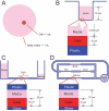
Media volumes in cell culture and organ constructs. A) A representation of the typical spherical volumes occupied by a single cell (~1 pL) and the media that is required to keep that cell viable for one day is a thousand times larger (~1 nL). B) Cells cultured in a well plate with daily media changes, wherein the volume of media is 1000 times the cell volume and hence the media height is approximately 10 mm, which results in a dilution of dynamic metabolites and paracrine and autocrine signals by a factor of one thousand. C) An organ construct grown in a microfluidic device with reservoirs that use gravity and height differences to superfuse the construct. The total system volume may lead to significant dilution of transient signaling molecules and metabolites. D) Coupled microphysiological systems in which each organ and the system’s fluid volume are scaled to the same functional size, and a low-volume, on-chip pump is used to recirculate the media and provide appropriate shear forces. The volume of the tubing and pump is comparable to the scaled, total human blood volume that includes both the vascular system and the missing organs. The mechanisms for delivery of O2 and nutrients and removal of CO2 and wastes are not shown. In (C) and (D), the cells will ideally be supported by an extracellular matrix that contains appropriate cell types to reflect the organ microenvironment and cellular heterogeneity.

References

    1. Martin HN. The direct influence of gradual variations of temperature upon the rate of beat of the dog’s heart. Philos Trans R Soc London. 1883 Jan 1;174:663–88.
    1. Langendorff O. Untersuchungen am Überlebenden Säugethierherzen. Pflug Arch Eur J Phy. 1895;61(6):291–332.
    1. Avis FR. Investigations of liver and kidney. Design of a dual apparatus for research study. Science Teacher. 1961;28(1):14–8.
    1. Neely JR, Liebermeister H, Battersby EJ, Morgan HE. Effect of pressure development on oxygen consumption by isolated rat heart. Am J Physiol. 1967 Apr 1;212(4):804–14. - PubMed
    1. Neely JR, Liebermeister H, Morgan HE. Effect of pressure development on membrane transport of glucose in isolated rat heart. Am J Physiol. 1967;212(4):815–22. - PubMed

Publication types