Review
. 2013 Mar;1(2):77-86.
doi: 10.1159/000346765.

Neuroimaging diagnosis and the collateral circulation in moyamoya disease

Affiliations
Review

Neuroimaging diagnosis and the collateral circulation in moyamoya disease

Wenhua Liu et al. Interv Neurol. 2013 Mar.

Abstract

Moyamoya disease (MMD) is an uncommon cerebrovascular disease that is characterized by progressive stenosis of the terminal portion of the internal carotid artery and its main branches, which is accompanied by the development of dilated, fragile collateral vessels at the base of the brain. This review will present different neuroimaging modalities for the diagnosis of MMD. Importantly, we will discuss the role of hyperintense vessels on fluid-attenuated inversion recovery images and their contribution to the evaluation of collateral patterns in MMD patients. Additionally, this review will summarize these common collateral patterns of MMD assessed by conventional cerebral angiography, and the associations of these angiographic collateral patterns with cerebrovascular lesions, including ischemia and hemorrhage will also be reviewed.

Keywords: Angiography; Collateral blood flow; Fluid-attenuated inversion recovery; Hyperintense vessels; Moyamoya disease.


Figures
Fig. 1
Fig. 1
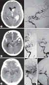
CT scans and angiograms showing neuroimaging characteristics of MMD patients. a CT scan of cerebral infarction and atrophy in the right territory of the MCA. b Angiogram of the terminal occlusion of the ICA with netlike vessels. c CT scan of a left caudate nucleus hematoma and a right anterior watershed zone. d, e Angiograms of the terminal occlusion of the ICA and the moyamoya vessels in the basal ganglia.
Fig. 2
Fig. 2
T2-weighted MRI modality showing neuroimaging characteristics of MMD patients. a The steno-occlusive lesion of the circle of Willis is well disclosed in axial T2-weighted images at the level of the basal cistern. b Enlarged image of the inset in a; the M1 segment of the MCA is not identified, and small signal voids corresponding to moyamoya vessels are visualized. c, d Angiograms showing the terminal occlusion of the ICA and the moyamoya vessels in the basal ganglia.
Fig. 3
Fig. 3
MRA modalities with different tesla scanners present neuroimaging characteristics of MMD patients. a A 0.5-tesla scanner shows a steno-occlusive lesion of the circle of Willis. b A 1.5-tesla MRA established a diagnosis of MMD with unclear intra- and extracranial collateral pathway. c A 3.0-tesla machine helps to detect some small moyamoya vessels and possible collateral pathways for MMD.
Fig. 4
Fig. 4
Progression of HV on FLAIR-MRI and transformation of ischemic lesion patterns on axial FLAIR images in an MMD patient. a Initial HV locate in territories of the anterior cerebral circulation, especially in regions of the MCA (arrowheads). HV are not seen obviously in PCA territories. There is a small subcortical infarct located in the territory of the anterior cerebral circulation (arrow). b Four months later, HV progression is prominent in territories of the anterior cerebral circulation (white arrowheads) compared with the initial HV image (a). A few HV are visible in territories of the PCA (black arrowheads). In addition, the subcortical infarct is transformed from the anterior to the posterior cerebral circulation (arrow). c Eight months later, many new HV locate in cerebral sulci or on the brain surface compared with previous images (a, b), being especially prominent in PCA territories (arrowheads). Moreover, the cerebral infarct pattern is shifted from the subcortical to the cortical area of the brain (arrow). d–f Bilateral carotid and right vertebral angiographies establish a diagnosis of MMD.
Fig. 5
Fig. 5
Conventional cerebral angiography demonstrates that the slow leptomeningeal retrograde blood flows are the origin of HV in an MMD patient. a HV on FLAIR within the bilateral territories of the sylvian fissure and the cerebral sulci of the temporal-occipital junction (frontal lobe). b Serial arteriographic images from a selective left ICA injection show the retrograde collateral flow via leptomeningeal branches of the PCA, corresponding to HV within the left territory of the sylvian fissure (short arrow). c, d Serial left vertebral angiograms demonstrate the leptomeningeal collateral vessels filling retrogradely the distal MCA, similarly corresponding to HV within the left territory of the sylvian fissure (long arrow).
Fig. 6
Fig. 6
A 3D reconstruction of a CT angiography showing an MMD patient.
Fig. 7
Fig. 7
Collateral patterns of MMD detected by conventional cerebral angiography. a ACA supplying the blood flow to the MCA territory via the MLA (arrow). b Dilation and extension of the AChA beyond the choroid fissure (arrow). c Patent PComA (arrow) providing blood flow to the territory of ACA and/or MCA via the PCA and MLA. d Blood flows from the posterior choroidal artery to the ACA territory via the posterior pericallosal artery (arrow). e Blood flows from the PCA to the ACA and/or MCA territory via the MLA (arrow). f Blood flows from the ophthalmic artery to the ACA territory via neovessels (arrow). g Middle MLA supplying blood to the ACA (white arrow) and/or MCA (black arrow). h Superficial temporal artery supplying blood to the MCA (arrow). i Occipital artery supplying blood to the ACA and/or MCA (arrows).
Fig. 8
Fig. 8
Association of hemorrhagic stroke with high-grade AChA-PComA. Patient 1: CT scan shows left intraventricular hemorrhage (a) and left ICA angiogram demonstrates grade 2 AChA-PComA (b). Patient 2: CT scan reveals temporal hemorrhage (c) and left ICA and vertebral artery angiograms show grade 2 AChA-PComA (d). Patient 3: CT scan reveals subarachnoid hemorrhage in the basal and ambient cistern (e); right vertebral artery angiogram demonstrates a large aneurysm (arrowhead) originating at the right bifurcation of the basilar artery (f), and right ICA angiogram shows grade 3 AChA-PComA (g).
Fig. 9
Fig. 9
Association of cerebral infarction with high-grade AChA-PComA. a Axial diffusion-weighted MR image demonstrating infarcted lesions in the territory of the left posterior circulation (arrows). b, c Right ICA angiograms disclose grade 2 AChA-PComA. d, e Left ICA angiograms show grade 3 AChA-PComA. f Left vertebral artery angiogram reveals collateral blood flow to the territory of the left MCA.

References

    1. Suzuki J, Takaku A. Cerebrovascular ‘moyamoya’ disease. Disease showing abnormal net-like vessels in base of brain. Arch Neurol. 1969;20:288–299. - PubMed
    1. Liu W, Zhu S, Wang X, Yue X, Zhou Zhi, Wang H, Xu G, Zhou C, Liu X. Evaluation of angiographic changes of the anterior choroidal and posterior communicating arteries for predicting cerebrovascular lesions in adult moyamoya disease. J Clin Neurosci. 2011;18:374–378. - PubMed
    1. Harada A, Fujii Y, Yoneoka Y, Takeuchi S, Tanaka R, Nakada T. High-field magnetic resonance imaging in patients with moyamoya disease. J Neurosurg. 2001;94:233–237. - PubMed
    1. Fukui M. Guidelines for the diagnosis and treatment of spontaneous occlusion of the circle of Willis (‘moyamoya’ disease). Research Committee on Spontaneous Occlusion of the Circle of Willis (Moyamoya Disease) of the Ministry of Health and Welfare, Japan. Clin Neurol Neurosurg. 1997;99(suppl 2):S238–S240. - PubMed
    1. Houkin K, Aoki T, Takahashi A, Abe H. Diagnosis of moyamoya disease with magnetic resonance angiography. Stroke. 1994;25:2159–2164. - PubMed