

Endovascular management of arteriovenous malformations of the brain
- PMID: 25187772
- PMCID: PMC4138962
- DOI: 10.1159/000346927
Endovascular management of arteriovenous malformations of the brain
Abstract
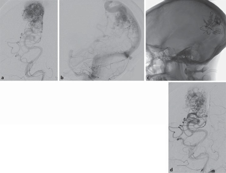
Arteriovenous malformations (AVMs) of the brain are rare, complex, vascular lesions that can result in significant morbidity and mortality. Modern treatment of brain AVMs is a multimodality endeavor, requiring a multidisciplinary team with expertise in cerebrovascular neurosurgery, endovascular intervention, and radiation therapy in order to provide all therapeutic options and determine the most appropriate treatment regimen depending on patient characteristics and AVM morphology. Current therapeutic options include microsurgical resection, radiosurgery (focused radiation), and endovascular embolization. Endovascular embolization is primarily used as a preoperative adjuvant before microsurgery or radiosurgery. Palliative embolization has been used successfully to reduce the risk of hemorrhage, alleviate clinical symptoms, and preserve or improve neurological function in inoperable or nonradiosurgical AVMs. Less frequently, embolization is used as 'primary therapy' particularly for smaller, surgically difficult lesions. Current embolic agents used to treat brain AVMs include both solid and liquid agents. Liquid agents including N-butyl cyanoacrylate and Onyx are the most commonly used agents. As newer embolic agents become available and as microcatheter technology improves, the role of endovascular treatment for brain AVMs will likely expand.
Keywords: Arteriovenous malformations; Embolization; Endovascular treatment; Microcatheters; Multimodality treatment.
Figures
References
-
- Richling B, Killer M, Al-Schameri AR, Ritter L, Agic R, Krenn M. Therapy of brain arteriovenous malformations: multimodality treatment from a balanced standpoint. Neurosurgery. 2006;59(suppl 3):S148–S157. - PubMed
-
- Morgan MK, Zurin AA, Harrington T, Little N. Changing role for preoperative embolisation in the management of arteriovenous malformations of the brain. J Clin Neurosci. 2000;7:527–530. - PubMed
-
- Taylor CL, Dutton K, Rappard G, Pride GL, Replogle R, Purdy PD, White J, Giller C, Kopitnik TA, Jr, Samson DS. Complications of preoperative embolization of cerebral arteriovenous malformations. J Neurosurg. 2004;100:810–812. - PubMed
-
- Ogilvy CS, Stieg PE, Awad I, Brown RD, Jr, Kondziolka D, Rosenwasser R, Young WL, Hademenos G, Special Writing Group of the Stroke Council, American Stroke Association AHA Scientific Statement. Recommendations for the management of intracranial arteriovenous malformations: a statement for healthcare professionals from a special writing group of the Stroke Council, American Stroke Association. Stroke. 2001;32:1458–1471. - PubMed
-
- Deruty R, Pelissou-Guyotat I, Mottolese C, Bascoulergue Y, Amat D. The combined management of cerebral arteriovenous malformations. Experience with 100 cases and review of the literature. Acta Neurochir (Wien) 1993;123:101–112. - PubMed
Publication types
LinkOut - more resources
Full Text Sources
Research Materials
