

An uncommon intramedullary tumor: primary spinal cord melanoma
- PMID: 25187871
- PMCID: PMC4149997
- DOI: 10.4184/asj.2014.8.4.512
An uncommon intramedullary tumor: primary spinal cord melanoma
Abstract
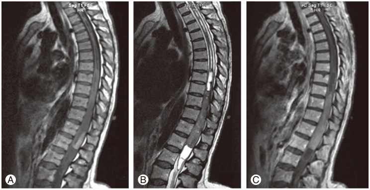
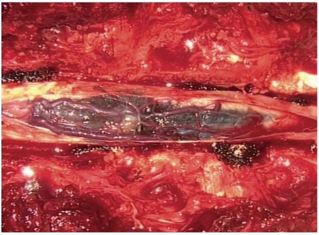
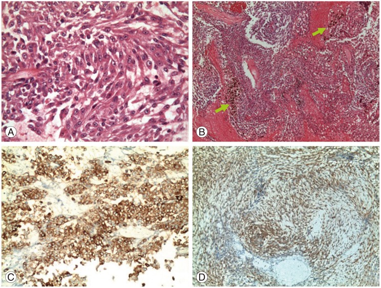
A 47-year-old woman was admitted with complaints of progressive weakness in the lower extremities and pain in the back and left leg. Thoracic magnetic resonance imaging (MRI) revealed a spinal intramedullary tumor between the T9 and L1 levels, which were iso- to hypointense on T2 and slightly hyperintense on T1-weighted images. The tumor was resected as total, and the diagnosis was malignant melanoma confirmed with histopathology. Neurological findings improved at the postoperative period and no residual or recurrence was noted on postoperative MRI at the 9-month follow-up. Primary melanoma of the spinal cord, particularly intramedullar localization, is seldomly reported in the literature. We report a primary malignant melanoma of the spinal cord and emphasize the diagnostic and prognostic challenges.
Keywords: Intramedullary; Malignant melanoma; Spinal cord neoplasms.
Conflict of interest statement
No potential conflict of interest relevant to this article was reported.
Figures
Similar articles
-
Primary intramedullary melanoma of lumbar spinal cord: A case report.World J Clin Cases. 2021 Apr 6;9(10):2352-2356. doi: 10.12998/wjcc.v9.i10.2352. World J Clin Cases. 2021. PMID: 33869613 Free PMC article.
-
Primary spinal melanoma: a case report and literature review.Chin Med J (Engl). 2012 Nov;125(22):4138-41. Chin Med J (Engl). 2012. PMID: 23158158 Review.
-
Primary spinal cord melanoma.J Korean Neurosurg Soc. 2010 Aug;48(2):157-61. doi: 10.3340/jkns.2010.48.2.157. Epub 2010 Aug 31. J Korean Neurosurg Soc. 2010. PMID: 20856666 Free PMC article.
-
Primary Spinal Melanoma With Intra- and Extradural Extensions: A Rare Case.Cureus. 2021 Jan 22;13(1):e12855. doi: 10.7759/cureus.12855. Cureus. 2021. PMID: 33633887 Free PMC article.
-
MR findings of a primary intramedullary malignant melanoma: case report and literature review.AJNR Am J Neuroradiol. 2001 Nov-Dec;22(10):1864-6. AJNR Am J Neuroradiol. 2001. PMID: 11733317 Free PMC article. Review.
Cited by
-
Primary spinal melanoma with special imaging appearances and pathologic characteristics: surgical treatment for two cases in a single center.Acta Neurol Belg. 2024 Apr;124(2):737-742. doi: 10.1007/s13760-023-02436-2. Epub 2023 Nov 23. Acta Neurol Belg. 2024. PMID: 37995022 No abstract available.
-
Primary Spinal Cord Melanoma of Intradural Extramedullary Origin.J Neurosci Rural Pract. 2019 Jul;10(3):522-525. doi: 10.1055/s-0039-1697559. Epub 2019 Oct 7. J Neurosci Rural Pract. 2019. PMID: 31595127 Free PMC article.
-
Primary Spinal Intradural Melanocytoma of the Thoracic Region: A Rare Case.Cureus. 2023 Jun 27;15(6):e41019. doi: 10.7759/cureus.41019. eCollection 2023 Jun. Cureus. 2023. PMID: 37519491 Free PMC article.
-
Clinical features and long-term outcomes of primary spinal malignant melanoma: a single center experience.J Neurooncol. 2017 Dec;135(3):513-519. doi: 10.1007/s11060-017-2593-7. Epub 2017 Aug 17. J Neurooncol. 2017. PMID: 28819705
-
Intramedullary primary spinal cord melanoma: illustrative case.J Neurosurg Case Lessons. 2025 Mar 10;9(10):CASE24732. doi: 10.3171/CASE24732. Print 2025 Mar 10. J Neurosurg Case Lessons. 2025. PMID: 40063999 Free PMC article.
References
-
- Fuld AD, Speck ME, Harris BT, et al. Primary melanoma of the spinal cord: a case report, molecular footprint, and review of the literature. J Clin Oncol. 2011;29:e499–e502. - PubMed
-
- Pappenheim E, Bhattacharji SK. Primary melanoma of the central nervous system. Clinical-pathological report of a case, with survey and discussion of the literature. Arch Neurol. 1962;7:101–113. - PubMed
-
- Larson TC, 3rd, Houser OW, Onofrio BM, Piepgras DG. Primary spinal melanoma. J Neurosurg. 1987;66:47–49. - PubMed
LinkOut - more resources
Full Text Sources
Other Literature Sources
