. 2014 Sep 4;9(9):e106487.
doi: 10.1371/journal.pone.0106487. eCollection 2014.

High density lipoprotein stimulated migration of macrophages depends on the scavenger receptor class B, type I, PDZK1 and Akt1 and is blocked by sphingosine 1 phosphate receptor antagonists

Affiliations

High density lipoprotein stimulated migration of macrophages depends on the scavenger receptor class B, type I, PDZK1 and Akt1 and is blocked by sphingosine 1 phosphate receptor antagonists

Aishah Al-Jarallah et al. PLoS One. .

Erratum in

Abstract

HDL carries biologically active lipids such as sphingosine-1-phosphate (S1P) and stimulates a variety of cell signaling pathways in diverse cell types, which may contribute to its ability to protect against atherosclerosis. HDL and sphingosine-1-phosphate receptor agonists, FTY720 and SEW2871 triggered macrophage migration. HDL-, but not FTY720-stimulated migration was inhibited by an antibody against the HDL receptor, SR-BI, and an inhibitor of SR-BI mediated lipid transfer. HDL and FTY720-stimulated migration was also inhibited in macrophages lacking either SR-BI or PDZK1, an adaptor protein that binds to SR-BI's C-terminal cytoplasmic tail. Migration in response to HDL and S1P receptor agonists was inhibited by treatment of macrophages with sphingosine-1-phosphate receptor type 1 (S1PR1) antagonists and by pertussis toxin. S1PR1 activates signaling pathways including PI3K-Akt, PKC, p38 MAPK, ERK1/2 and Rho kinases. Using selective inhibitors or macrophages from gene targeted mice, we demonstrated the involvement of each of these pathways in HDL-dependent macrophage migration. These data suggest that HDL stimulates the migration of macrophages in a manner that requires the activities of the HDL receptor SR-BI as well as S1PR1 activity.


Conflict of interest statement

Competing Interests: The authors have declared that no competing interests exist.

Figures

Figure 1
Figure 1. HDL stimulates the migration of macrophages but not of lipid loaded macrophage foam cells.
A. Wild type mouse peritoneal macrophages were incubated in lipoprotein deficient serum overnight and cell migration in response to no stimulus (control), apoA1(100 µg/ml), HDL (100 µg protein/ml) or MCP-1 (100 ng/ml) (each added to the bottom well of the migration assay chamber) was performed as described in the Methods section. The number of migrated cells/well from three independent samples from each group is represented as the mean ± standard deviation. B. Wild type mouse peritoneal macrophages, or C. RAW 264.7 cells were incubated in collagen I coated cell culture dishes with either HDL (100 µg protein/ml), FTY720 (2 ng/ml) or MCP-1 (100 ng/ml) under conditions paralleling the migration assay. Cell adhesion was measured by counting DAPI stained nuclei. The degree of cell adhesion was normalized to that in control cells and is represented as the mean ± standard deviation of triplicates. D. Foam cells from wild type mouse peritoneal macrophages were generated by culture for 48 hrs in the presence of AcLDL (100 µg/ml). Cells were washed and migration in response to HDL was measured as described in the Methods section. The number of migrated cells/well is plotted as the mean ± standard deviation of triplicates. Statistical analysis was done using one way ANOVA with Holm-Sidak post hoc test (A–C) or Student's T-test (D). Values identified with different letters are statistically significantly different (P<0.05). NS indicates that no statistically significant difference was detected.
Figure 2
Figure 2. HDL-induced migration is reduced in macrophages deficient in either SR-BI or PDZK1.
A. Peritoneal macrophages were prepared from either wild type (WT) or SR-BI KO mice, and cultured in the presence of 3% NCLPDS for 16 hrs before analysis. Migration of cells in response to no stimulus (control), HDL (100 µg protein/ml), FTY720 (2 ng/ml) or MCP-1 (100 ng/ml) as described in the legend to Figure 1 and the Methods section. B. Peritoneal macrophages from WT or SR-BI KO mice were cultured as described in A, prior to incubation with or without HDL (100 µg protein/ml) for the times indicated. Actin filaments were visualized by fluorescence microscopy after alexa 488-phalloidin staining. Representative images are shown. Scale bars = 10 µm. C. Numbers of cells with lamellipodia (arrows in B) were counted (∼100 cells over 4–5 fields) for cells isolated from three mice from each genotype. D. Migration of peritoneal macrophages from WT or PDZK1 KO mice as described for panel A. E. Flow cytometry analysis of cell surface SR-BI levels in wild type, SR-BI KO and PDZK1 KO macrophages. Values in A, C and D are means ± standard deviations of triplicates and are representative of multiple independent assays. Data in E is representative of multiple analyses. Statistical analysis in panels A, C and D was done using two way ANOVA with Tukey post hoc test. Values identified with different letters are statistically significantly different (P<0.001). NS indicates that no statistically significant difference was detected.
Figure 3
Figure 3. Antibody or small molecule mediated inactivation of SR-BI inhibits HDL dependent but not FTY720 dependent macrophage migration.
Mouse peritoneal macrophages were pretreated for 30 min with either A. An anti-SR-BI blocking antiserum or pre-immune rabbit serum (0.5 µg/ml); or B. BLT-1 (0.3 µM), an inhibitor of SR-BI mediated lipid transfer, or DMSO vehicle control. Cell migration in response to HDL (100 µg protein/ml), FTY720 (2 ng/ml) or MCP-1 (100 ng/ml) was measured in the continued presence of the inhibitors as described in the legend to Figure 1 and the Methods section. Data are means ± standard deviations of 6 replicates. Statistical analysis was done using two way ANOVA with Tukey post hoc test. Values identified with different letters are statistically significantly different (P<0.002).
Figure 4
Figure 4. Blockade of S1PR1 prevents HDL stimulated macrophage migration.
RAW 264.7 (panels A,B) or mouse peritoneal macrophages (panel D) were pretreated for 30 min with the following: A. PTX (100 ng/ml) an inhibitor of Gαi protein coupled receptors; B. VPC23019 (10 µM) an antagonist of S1PR's 1, and 3; or D. W-146 (10 µM) an antagonist specific for S1PR1. Control cells were treated with either 2 µg/ml BSA or DMSO vehicle as indicated. Cell migration in response to HDL (100 µg protein/ml), FTY720 (2 ng/ml), MCP-1 (100 ng/ml) and/or SEW2871 (5 nM) was measured in the continued absence or presence of the inhibitors as described in the legend to Figure 1 and the Methods section. C. Analysis of expression of S1PR1, 2 and 3 in peritoneal macrophages (left panel) or lung tissue (right panel) from wild type mice. Data are means ± standard deviations of 6 replicates (A,B) or 3 replicates (C,D). Statistical analysis was done using two way ANOVA with Tukey post hoc test. Values identified with different letters are statistically significantly different (P<0.002).
Figure 5
Figure 5. HDL stimulated macrophage migration involves Rho kinase and PI3K-Akt 1 signaling.
A. RAW 264.7 cells were cultured in media containing 3% NCLPDS for 18 hrs. Cells were pre-incubated with 10 µM of either the Rho Kinase inhibitor, Y-27632, or the PI3K inhibitor LY294002, or with DMSO vehicle for 30 min and then the migration in response to HDL (100 µg protein/ml), FTY720 (2 ng/ml) or MCP-1 (100 ng/ml) was measured in the continued presence or absence of the indicated inhibitors. B. RAW 264.7 cells were serum starved for 18 hrs, washed and treated with or without HDL (100 µg protein/ml) for 10, 30 or 60 min. Equal amounts of proteins were analyzed by SDS-PAGE and immunoblotting for either phospho-Ser473- or total-Akt. C. MPM's were harvested from 6 independent WT, Akt1 KO, Akt2 KO or Akt3 KO mice. Migration in response to no stimulus, HDL (100 µg protein/ml), FTY720 (2 ng/ml) or MCP-1 (100 ng/ml) was measured. Data from A and C are means ± standard deviations of 6 replicates done over two independent experiments. Values identified with different letters are statistically significantly different (P<0.003, 2 way ANOVA with Tukey post hoc test). D. Flow cytometry analysis of cell surface SR-BI levels in wild type, SR-BI KO and Akt1 KO macrophages. Shown are representative histograms of an experiment performed twice.
Figure 6
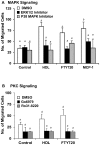
Figure 6. HDL stimulated macrophage migration involves MAP kinase and PKC pathways.
RAW 264.7 cells were cultured in media containing 3%NCLPDS for 18 hrs. Cells were pre-incubated with the indicated inhibitors for 30 min and then the migration in response to HDL (100 µg protein/ml), FTY720 (2 ng/ml) or MCP-1 (100 ng/ml) was measured in the continued presence or absence of the indicated inhibitors. Control cells were treated with DMSO vehicle. A. Cells were treated with either 10 µM of the ERK1/2 pathway inhibitor, PD98059, or 1 µM of the p38 MAPK inhibitor, SB230580. B. Cells were treated with 5 µM of the PKC inhibitors Ro31-8220 or Go6976. Data are means ± standard deviations of 3 replicates. Values identified with different letters are statistically significantly different (P<0.04, 2 way ANOVA with Tukey post hoc test).
Figure 7
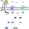
Figure 7. Working model for HDL mediated stimulation of macrophage migration.
HDL binding to SR-BI leads to activation of S1PR1 signaling. This may involve transfer of S1P from bound HDL to S1PR1. Inhibition of HDL binding to SR-BI (blocking antibody) or SR-BI-mediated lipid transfer (BLT-1) prevents HDL dependent activation of S1PR1 signaling, but does not affect direct activation of S1PR1 by agonists. Inactivation of expression of SR-BI or PDZK1, on the other hand, inhibits migratory responses to FTY720, through an as yet unknown mechanism. HDL dependent activation of migration is suppressed by inhibition of S1PR1 signaling with FTY720 or W146, which directly antagonize S1PR1, or with PTX, which blocks Gαi coupled GPCR's including CCR2 (receptor for MCP-1). Upon appropriate stimulation, S1PR1 (and CCR2) stimulate macrophage migration by activation of diverse signaling pathways including PI3K/Akt1, Rho kinase, PKC, p38 MAPK and Erk1/2 pathways .

References

    1. Taylor PR, Martinez-Pomares L, Stacey M, Lin HH, Brown GD, et al. (2005) Macrophage receptors and immune recognition. Annu Rev Immunol 23: 901–944. - PubMed
    1. Libby P (2002) Inflammation in atherosclerosis. Nature 420: 868–874. - PubMed
    1. Steinberg D (1997) Low density lipoprotein oxidation and its pathobiological significance. J Biol Chem 272: 20963–20966. - PubMed
    1. Tabas I, Williams KJ, Boren J (2007) Subendothelial lipoprotein retention as the initiating process in atherosclerosis: update and therapeutic implications. Circulation 116: 1832–1844. - PubMed
    1. Nagao T, Qin C, Grosheva I, Maxfield FR, Pierini LM (2007) Elevated cholesterol levels in the plasma membranes of macrophages inhibit migration by disrupting RhoA regulation. Arterioscler Thromb Vasc Biol 27: 1596–1602. - PubMed

Publication types

MeSH terms