. 2014 Sep 4;9(9):e107000.
doi: 10.1371/journal.pone.0107000. eCollection 2014.

Crystal structure of the mineralocorticoid receptor DNA binding domain in complex with DNA

Affiliations

Crystal structure of the mineralocorticoid receptor DNA binding domain in complex with DNA

William H Hudson et al. PLoS One. .

Abstract

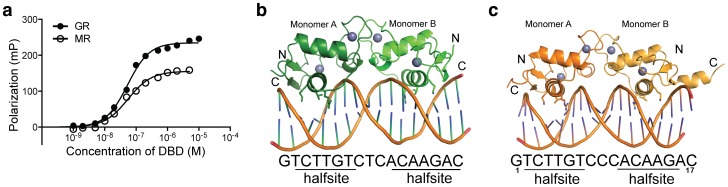
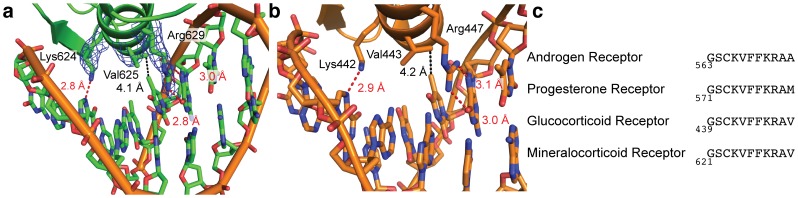
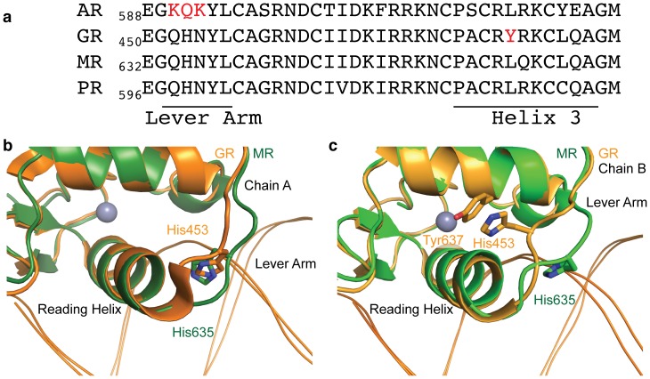
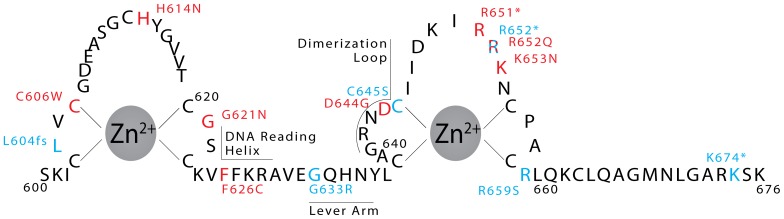
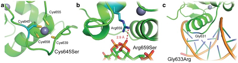
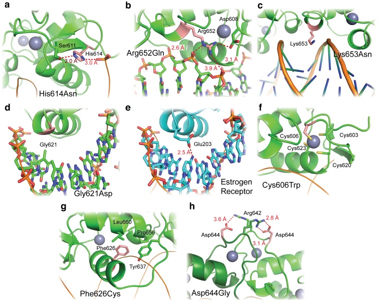
The steroid hormone receptors regulate important physiological functions such as reproduction, metabolism, immunity, and electrolyte balance. Mutations within steroid receptors result in endocrine disorders and can often drive cancer formation and progression. Despite the conserved three-dimensional structure shared among members of the steroid receptor family and their overlapping DNA binding preference, activation of individual steroid receptors drive unique effects on gene expression. Here, we present the first structure of the human mineralocorticoid receptor DNA binding domain, in complex with a canonical DNA response element. The overall structure is similar to the glucocorticoid receptor DNA binding domain, but small changes in the mode of DNA binding and lever arm conformation may begin to explain the differential effects on gene regulation by the mineralocorticoid and glucocorticoid receptors. In addition, we explore the structural effects of mineralocorticoid receptor DNA binding domain mutations found in type I pseudohypoaldosteronism and multiple types of cancer.


Conflict of interest statement

Competing Interests: The authors have declared that no competing interests exist.

Figures

Figure 1
Figure 1. Structure of the human mineralocorticoid DNA binding domain in complex with a glucocorticoid response element.
(a) The MR DBD binds to a GRE with approximately the same affinity as the GR DBD. (b) Overall structure of the MR DBD (green) bound to a 17 base pair GRE. The sequence of the element, along with the two bound half sites, is shown below the structure. In both panels (b) and (c), the structure shown depicts the asymmetric unit of the crystal structure and separate GR monomers are differentially colored. (c) Structure of the GR DBD (orange) bound to a similar GRE, with sequence and half sites indicated below. Panel (c) is derived from the structure of the GR DBD bound to the FKBP5 GRE, PDB 3G6P .
Figure 2
Figure 2. Sequence-specific DNA recognition by the MR DBD.
(a) Three residues mediate sequence-specific contacts by the MR DBD DNA reading helix. Lysine 624 makes a hydrogen bond with a guanine base, valine 625 makes van der Waals contacts with a thymine base, and arginine 629 makes two interactions with a guanine base. Electron density (composite omit 2Fo−Fc map with simulated annealing, contoured to 1 σ) is shown for the three protein side chains. (b) GR recognizes GREs in an identical manner as the MR DBD, using lysine 442, valine 443, and arginine 447 to contact analogous bases. (c) Sequence alignment showing conservation of the DNA reading helix among the NR3C receptors.
Figure 3
Figure 3. Lever arm conformation differs between MR and GR.
(a) Sequence alignment of the lever arm through helix 3 of the NR3C receptors. AR and GR contain divergent sequence at the lever arm and helix 3, respectively (red). In a dimer of GR molecules on DNA (see Figure 1c), the side chain of histidine 453 can assume two conformations. (b) Monomer A of the GR – FKBP5 GRE complex contains histidine 453 in a “flipped” conformation, where the histidine side chain sits between the DNA and DBD reading helix; a similar conformation is seen in monomer A of the MR DBD – GRE complex. (c) However, histidine 453 in the second GR DBD monomer assumes a “packed” position against tyrosine 637 in the core of the GR DBD fold. This conformation does not occur in the MR DBD – GRE structure, likely due to the presence of a leucine rather than tyrosine at position 660 (GR position 478). In panels (b) and (c), DNA is shown as a ribbon helix below the protein.
Figure 4
Figure 4. Analysis of the MR – DNA interface.
(a) Thermal motion of GREs when bound to GR (left) and MR (right). Thicker, red sections of DNA indicate higher B-factors and therefore higher thermal motion. (b) Major groove width at each position of the GRE when bound to the MR and GR DBDs. (c).
Figure 5
Figure 5. MR DBD mutations found in disease.
Mutations found in type I pseudohypoaldosteronism are in blue and mutations found in cancer are in red. An asterisk indicates a nonsense mutation, and fs indicates a frameshift mutation.
Figure 6
Figure 6. MR DBD mutations driving PHA1.
Mutated residues are shown in blue. (a) Cysteine 645 is one of four cysteines that coordinate a Zn2+ ion in MR’s second zinc finger. Its mutation to serine would destroy the zinc finger fold of the DBD. (b) Arginine 659 makes non-specific interactions with the DNA backbone, and is mutated to serine in some cases of PHA1. (c) Glycine 633 is part of the DBD lever arm, which is important for receptor activation . Mutation of this residue to arginine affects receptor activation without affecting its affinity for DNA .
Figure 7
Figure 7. MR DBD mutations found in cancer.
Mutated residues are shown in red. (a) Histidine 614 interacts with both the DNA backbone and serine 611. (b) Similarly, arginine 652 also interacts with the DNA backbone and a neighboring amino acid, aspartic acid 608. (c) Lysine 653 is within an appropriate distance to make non-specific contacts with the minor groove of a GRE. (d) Glycine 621 is mutated to aspartic acid in a stomach cancer sample. In the estrogen receptor, the homologous amino acid is the similar glutamic acid, which participates in base-specific DNA recognition (panel e). (f) Cysteine 606 is one of four cysteine residues to coordinate a Zn2+ ion in one of MR’s two zinc fingers. (g) Phenylalanine 626 comprises part of the hydrophobic core of the DBD. (h) Aspartic acid 644 is a key mediator of MR dimerization, forming a salt bridge with arginine 642 of the second monomer.

References

    1. Falkenstein E, Tillmann HC, Christ M, Feuring M, Wehling M (2000) Multiple actions of steroid hormones–a focus on rapid, nongenomic effects. Pharmacol Rev 52: 513–556. - PubMed
    1. Tsai M, O’Malley BW (1994) Molecular mechanisms of action of steroid/thyroid receptor superfamily members. Annu Rev Biochem 63: 451–486. - PubMed
    1. Rastinejad F, Huang P, Chandra V, Khorasanizadeh S (2013) Understanding nuclear receptor form and function using structural biology. J Mol Endocrinol 51: T1–T21. - PMC - PubMed
    1. Bridgham JT, Carroll SM, Thornton JW (2006) Evolution of hormone-receptor complexity by molecular exploitation. Science 312: 97–101. - PubMed
    1. Marver D, Stewart J, Funder JW, Feldman D, Edelman IS (1974) Renal aldosterone receptors: studies with (3H)aldosterone and the anti-mineralocorticoid (3H)spirolactone (SC-26304). Proc Natl Acad Sci U S A 71: 1431–1435. - PMC - PubMed

Publication types

MeSH terms