

Investigating the role of free-living amoebae as a reservoir for Mycobacterium ulcerans
- PMID: 25188535
- PMCID: PMC4154674
- DOI: 10.1371/journal.pntd.0003148
Investigating the role of free-living amoebae as a reservoir for Mycobacterium ulcerans
Abstract
Background: The reservoir and mode of transmission of Mycobacterium ulcerans, the causative agent of Buruli ulcer, still remain a mystery. It has been suggested that M. ulcerans persists with difficulty as a free-living organism due to its natural fragility and inability to withstand exposure to direct sunlight, and thus probably persists within a protective host environment.
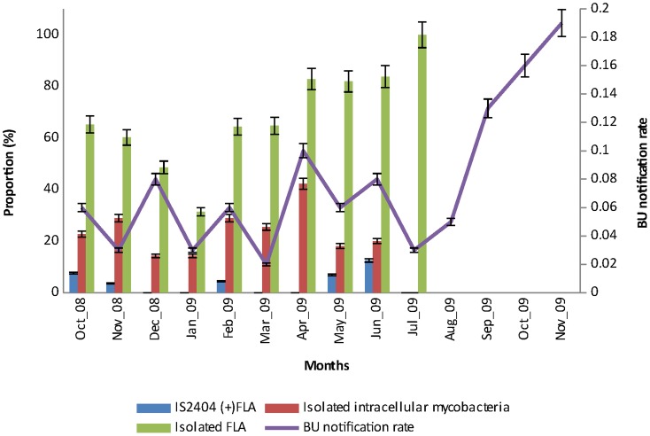
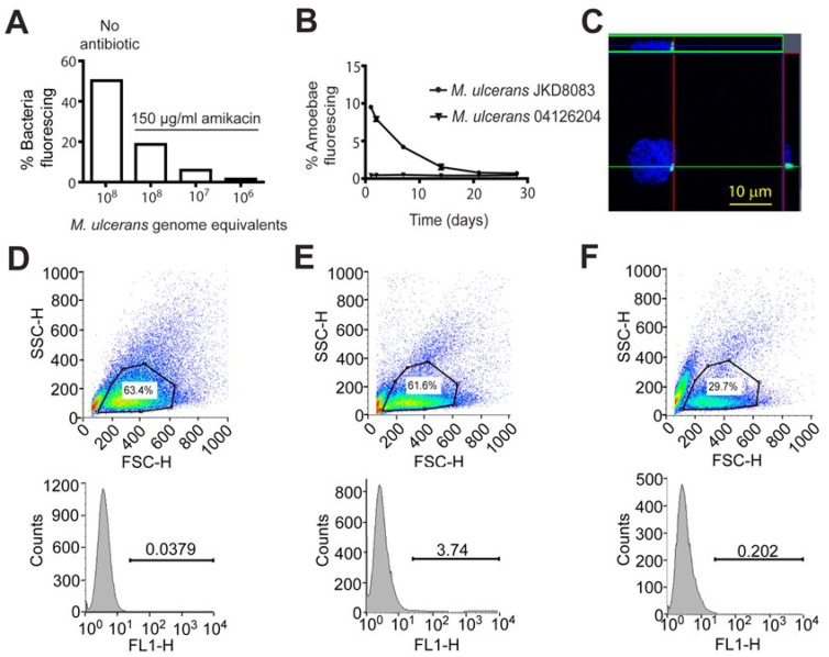
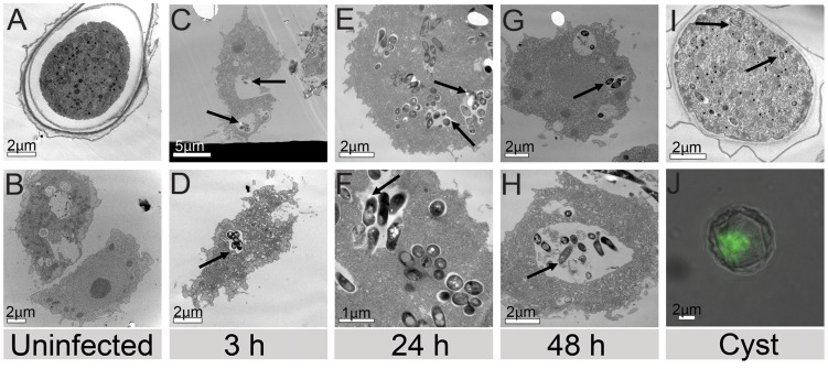
Methodology/principal findings: We investigated the role of free-living amoebae as a reservoir of M. ulcerans by screening the bacterium in free-living amoebae (FLA) cultures isolated from environmental specimens using real-time PCR. We also followed the survival of M. ulcerans expressing green fluorescence protein (GFP) in Acanthameoba castellanii by flow cytometry and observed the infected cells using confocal and transmission electron microscopy for four weeks in vitro. IS2404 was detected by quantitative PCR in 4.64% of FLA cultures isolated from water, biofilms, detritus and aerosols. While we could not isolate M. ulcerans, 23 other species of mycobacteria were cultivated from inside FLA and/or other phagocytic microorganisms. Laboratory experiments with GFP-expressing M. ulcerans in A. castellani trophozoites for 28 days indicated the bacteria did not replicate inside amoebae, but they could remain viable at low levels in cysts. Transmission electron microscopy of infected A. castellani confirmed the presence of bacteria within both trophozoite vacuoles and cysts. There was no correlation of BU notification rate with detection of the IS2404 in FLA (r = 0.07, n = 539, p = 0.127).
Conclusion/significance: This study shows that FLA in the environment are positive for the M. ulcerans insertion sequence IS2404. However, the detection frequency and signal strength of IS2404 positive amoabae was low and no link with the occurrence of BU was observed. We conclude that FLA may host M. ulcerans at low levels in the environment without being directly involved in the transmission to humans.
Conflict of interest statement
The authors have declared that no competing interests exist.
Figures

References
-
- Portaels F, Silva MT, Meyers WM (2009) Buruli ulcer. Clin Dermatol 27: 291–305. - PubMed
-
- Marion E, Deshayes C, Chauty A, Cassisa V, Tchibozo S, et al. (2011) Detection of Mycobacterium ulcerans DNA in water bugs collected outside the aquatic environment in Benin. Med Trop (Mars) 71: 169–172. - PubMed
-
- Merritt RW, Walker ED, Small PLC, Wallace JR, Johnson PDR, et al. (2010) Ecology and transmission of buruli ulcer disease: a systematic review. PLoS Negl Trop Dis 4: e911 Available: http://www.pubmedcentral.nih.gov/articlerender.fcgi?artid=3001905&tool=p.... Accessed 4 May 2014. - PMC - PubMed
Publication types
MeSH terms
LinkOut - more resources
Full Text Sources
Other Literature Sources
