. 2014 Oct;9(10):2329-40.
doi: 10.1038/nprot.2014.158. Epub 2014 Sep 4.

Generation of cerebral organoids from human pluripotent stem cells

Affiliations

Generation of cerebral organoids from human pluripotent stem cells

Madeline A Lancaster et al. Nat Protoc. 2014 Oct.

Abstract

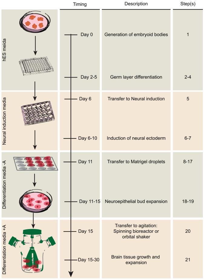
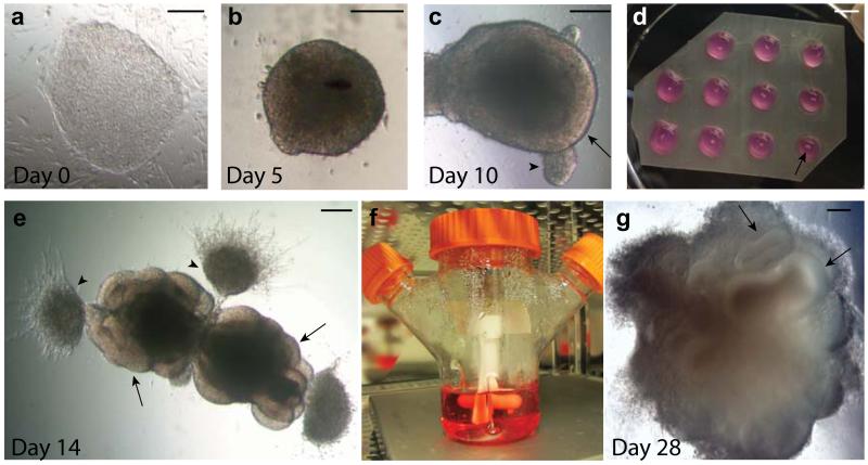
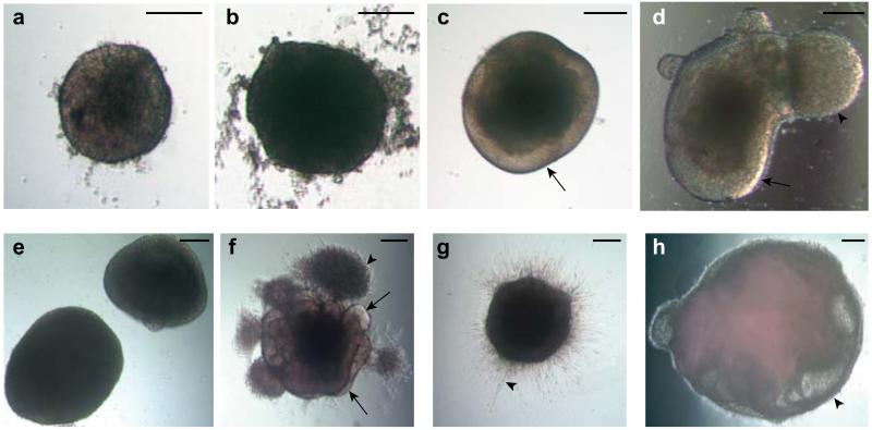
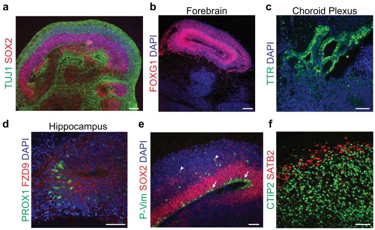
Human brain development exhibits several unique aspects, such as increased complexity and expansion of neuronal output, that have proven difficult to study in model organisms. As a result, in vitro approaches to model human brain development and disease are an intense area of research. Here we describe a recently established protocol for generating 3D brain tissue, so-called cerebral organoids, which closely mimics the endogenous developmental program. This method can easily be implemented in a standard tissue culture room and can give rise to developing cerebral cortex, ventral telencephalon, choroid plexus and retinal identities, among others, within 1-2 months. This straightforward protocol can be applied to developmental studies, as well as to the study of a variety of human brain diseases. Furthermore, as organoids can be maintained for more than 1 year in long-term culture, they also have the potential to model later events such as neuronal maturation and survival.


Figures

Figure 1
Figure 1. Schematic diagram of cerebral organoid method and timing
The protocol begins with the generation of EBs from human PSCs in a 96-well U-bottom plate. The day EBs are made is day 0. Generation of EBs is outlined in step 1. Feeding and monitoring the EBs is described in Steps 2-4. Typically on day 6 the EBs are transferred to a 24-well plate containing Neural induction media as described in step 5. Feeding and monitoring of neural induction is described in steps 6-7. On day 11, neuroectodermal tissues are then transferred to droplets of Matrigel on a sheet of dimpled Parafilm, as described in steps 8-17, and then grown in a 6 cm dish. Monitoring of these tissues is described in steps 18-19. Finally, Matrigel droplets are transferred to the spinning bioreactor on day 15, as described in step 20 and further maintained as described in step 21.
Figure 2
Figure 2. Progression of cerebral organoid development from human PSCs
a. A colony of feeder-dependent human ESCs showing typical pluripotent morphology with clear boundaries and a uniform texture. b. An EB at day 5 showing evidence of ectodermal differentiation as indicated by the presence of brightened surface tissue, whereas the center is quite dark with dense non-ectodermal tissue. The EB also has a smooth surface indicating healthy tissue. c. An early organoid at day 10 showing smooth edges and bright optically translucent surface tissue consistent with neuroectoderm (arrow). This organoid also contains small buds of ectodermal tissue that is not organized radially (arrowhead). d. Image of the neuroectodermal tissues embedded in Matrigel droplets on a sheet of dimpled Parafilm. The tissues are visible as small white specks within the droplet (arrow). e. An organoid at day 14, after embedding in Matrigel, showing evidence of neuroepithelial bud outgrowth (arrows) that are optically clear and in several cases surround a visible lumen. Other outgrowths and migrating cells are also visible (arrowheads) that are not neuroepithelial. f. An image of the spinning bioreactor setup in the tissue culture incubator. Organoids are visible within the bioreactor as small white floating specks. g. An organoid at day 28 of the protocol revealing many large neural tissues (arrows) that have greatly expanded once embedded in the Matrigel. Scale bar in a-c, e, g is 200 μm, and is 5 mm in d.
Figure 3
Figure 3. Examples of suitable and suboptimal organoids at various stages
a. An example of an optimal EB at day 5 showing brightening and clearing around the surface and with smooth edges. b. An example of an unsuitable EB lacking optical clearing and with large amounts of cell debris, despite its large size. c. An organoid in Neural induction media showing clear radially organized optically translucent neuroectoderm (arrow). d. An example of an acceptable organoid also showing evidence of optically clear neuroectoderm (arrow) but also a large bud of translucent ectoderm that is not radially organized (arrowhead). This bud, although not ideal, will not effect development of the neighboring neuroectodermal tissue. e. An example of failed neural induction. The EBs are too large and lack optically translucent, radially organized neuroectoderm. f. An ideal organoid soon after Matrigel embedding showing many buds of neuroepithelium (arrows) as well as non neuroepithelial cells which have migrated into the Matrigel (arrowhead). g. An organoid which has failed to produce neuroepithelial buds, instead displaying extended cell processes (arrowhead) consistent with direct neural differentiation. h. An example of a failed organoid after several weeks of differentiation showing large fluid-filled cysts that lack a thickened neuroepithelium (arrowhead). Scale bar is 200 μm in all panels.
Figure 4
Figure 4. Staining for brain regions and neuronal cell identities
a. Staining for neurons (TUJ1, green) and progenitors (SOX2, red) in a large continuous cortical tissue within an organoid. Note the organized apical progenitor zone surrounded by basally located neurons. b. A forebrain region of an organoid staining positive for the marker FOXG1 (red). c. Choroid plexus stains positive for the marker TTR (green) and displays convoluted cuboidal epithelium. d. Hippocampal regions stain positive for the markers PROX1 (green) and FZD9 (red), although the cells fail to spatially organize into recognizable dentate gyrus and CA regions. e. Staining for mitotic radial glia (P-Vimentin, P-Vim, green) in a cortical region reveals inner radial glia undergoing mitosis at the apical membrane (arrows), while outer radial glia undergo mitosis outside the ventricular zone (arrowheads). All radial glia are marked by SOX2 (red). f. Staining for cortical layer identities of advanced organoids (75 days). Later-born superficial layer identity (SATB2, red) neurons populate more superficial regions of the organoid, while early-born deep layer identity (CTIP2, green) neurons populate deeper regions of the organoid. DAPI in a-e labels nuclei (blue). Samples in a-e are 30-35 days after initiation of the protocol. Scale bar is 100 μm in a-b and 50 μm in c-f.

Comment in

  • Findings of Research Misconduct.
    [No authors listed] [No authors listed] Fed Regist. 2020 Aug 14;85(158):49661-49662. Fed Regist. 2020. PMID: 32831428 Free PMC article. No abstract available.

References

    1. Lanza R, Gearhart J, Hogan B, Melton D, Pedersen R. Essentials of stem cell biology. Elsevier. 2009
    1. Sato T, Clevers H. Growing self-organizing mini-guts from a single intestinal stem cell: mechanism and applications. Science. 2013;340:1190–1194. - PubMed
    1. Sasai Y, Eiraku M, Suga H. In vitro organogenesis in three dimensions: self-organising stem cells. Development. 2012;139:4111–4121. - PubMed
    1. Eiraku M, et al. Self-organizing optic-cup morphogenesis in three-dimensional culture. Nature. 2011;472:51–56. - PubMed
    1. Sato T, et al. Single Lgr5 stem cells build crypt-villus structures in vitro without a mesenchymal niche. Nature. 2009;459:262–265. - PubMed

Publication types

MeSH terms