Review
. 2014 Dec;96(6):672-83.
doi: 10.1038/clpt.2014.174. Epub 2014 Sep 4.

Serendipity and the discovery of novel compounds that restore mitochondrial plasticity

Affiliations
Review

Serendipity and the discovery of novel compounds that restore mitochondrial plasticity

H H Szeto et al. Clin Pharmacol Ther. 2014 Dec.

Abstract

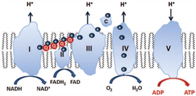
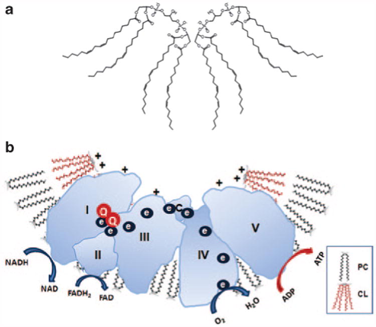
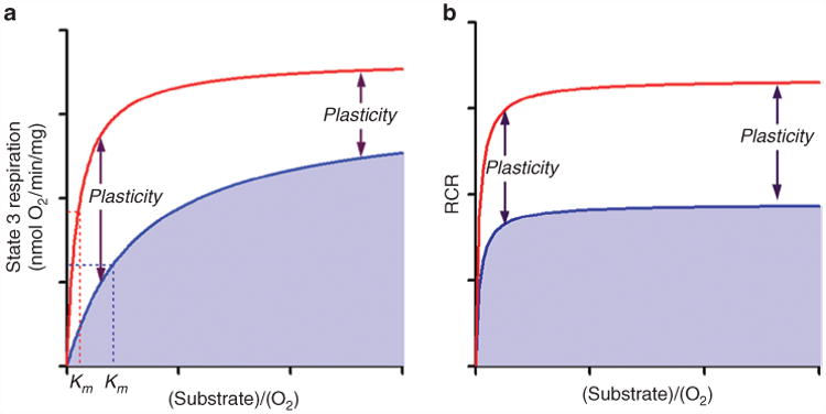
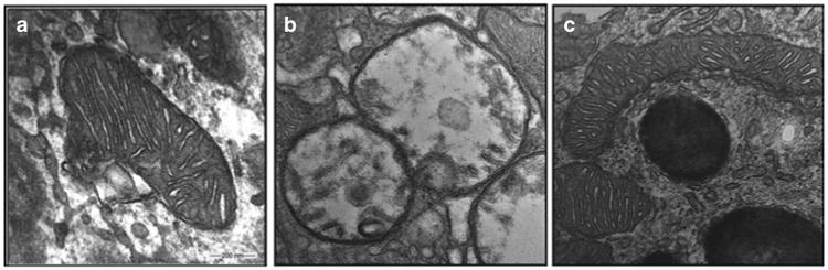
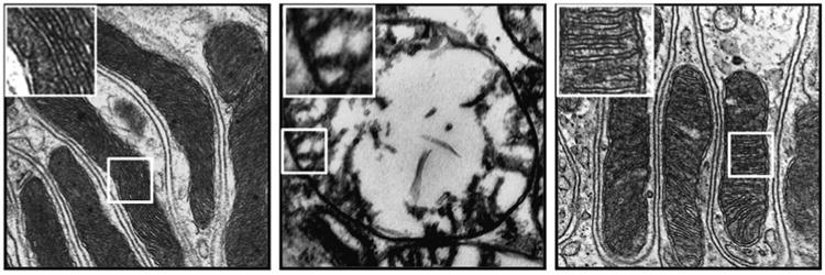
The mitochondrial electron transport chain (ETC) plays a central role in energy generation in the cell. Mitochondrial dysfunctions diminish adenosine triphosphate (ATP) production and result in insufficient energy to maintain cell function. As energy output declines, the most energetic tissues are preferentially affected. To satisfy cellular energy demands, the mitochondrial ETC needs to be able to elevate its capacity to produce ATP at times of increased metabolic demand or decreased fuel supply. This mitochondrial plasticity is reduced in many age-associated diseases. In this review, we describe the serendipitous discovery of a novel class of compounds that selectively target cardiolipin on the inner mitochondrial membrane to optimize efficiency of the ETC and thereby restore cellular bioenergetics in aging and diverse disease models, without any effect on the normal healthy organism. The first of these compounds, SS-31, is currently in multiple clinical trials.


Conflict of interest statement

Conflict of Interest: The SS peptides described in this article have been licensed for commercial research and development to Stealth Peptides, a clinical-stage biopharmaceutical company in which H.H.S., A.V.B., and the Cornell Research Foundation have financial interests. H.H.S. is the scientific founder of Stealth Peptides International and a consultant to Stealth Peptides. The Research Program in Mitochondrial Therapeutics was established at the Weill Cornell Medical College with a gift from Stealth Peptides International.

Figures

Figure 1
Figure 1
Mitochondria are composed of two membranes: an outer mitochondrial membrane and an inner mitochondrial membrane (IMM). The IMM is unique in that it has a very high concentration of proteins and is rich in cardiolipin, a very anionic phospholipid that plays a crucial role in the formation of cristae curvature and is important for efficient function of the electron transport chain (ETC). The mitochondrial matrix enclosed by the IMM contains not only mitochondrial DNA but also the major enzymes of the tricarboxylic acid cycle, which provide reducing equivalents in the form of NADH and FADH2 to the ETC. The IMM is compartmentalized into numerous cristae that greatly increase the surface area of the IMM. The four power-generating protein complexes of the ETC (complexes I–IV) reside on these cristae in the IMM, and the proton gradient generated across the IMM as a result of electron transfer from complex I to complex IV drives the production of ATP by the F0F1–ATPase (complex V). All these complexes must be assembled properly for efficient electron transfer to take place. ADP, adenosine diphosphate; ATP, adenosine triphosphate; ATPase, adenosine triphosphatase; FADH2, reduced flavin adenine dinucleotide; NADH, reduced nicotinamide adenine dinucleotide.
Figure 2
Figure 2
Cardiolipin promotes membrane curvature to optimize electron transport chain. (a) Cardiolipin (CL) is a dimeric phospholipid with a small acidic head group and four acyl chains, thus giving it a conical structure. As a result of its conical shape, CL exerts lateral pressure on a membrane containing other phospholipids such as phosphatidylcholine (PC), and results in membrane curvature. (b) CL rafts on cristae membranes allow the respiratory complexes to form supercomplexes that reduce the distance between redox partners. Besides providing a platform for the aggregation of the respiratory complexes, CL is also thought to serve as a proton trap on the outer leaflet of the inner mitochondrial membrane to allow rapid lateral diffusion of protons to the ATP synthase with minimal changes in the bulk phase pH. ADP, adenosine diphosphate; ATP, adenosine triphosphate; FADH2, reduced flavin adenine dinucleotide; NADH, reduced nicotinamide adenine dinucleotide.
Figure 3
Figure 3
The mitochondrial electron transport chain needs to be able to elevate its capacity to produce ATP at times of increased metabolic demand or decreased fuel supply. Its ability to do so has been called “spare respiratory capacity” or “mitochondrial plasticity.” (a) Mitochondrial state 3 respiration (ADP-stimulated respiration) is dependent on substrate concentration and O2 supply (blue line). Mitochondrial plasticity provides a shift in the Michaelis–Menten constant (Km) so that higher mitochondrial respiration can be achieved with lower substrate or O2 concentration (red line). The effect is less at high concentrations of substrates. (b) The respiratory control ratio (RCR) shows that state 4 respiration is not changed. Mitochondrial plasticity is determined by the efficacy of mitochondrial coupling between O2 consumption and the rate of ATP synthesis (P/O ratio). ADP, adenosine diphosphate; ATP, adenosine triphosphate.
Figure 4
Figure 4
SS-31 protects the mitochondrial structure in retinal pigment epithelium (RPE) cells in diabetic mice. Mice were fed either a normal diet (ND) or a diabetic diet (DD) starting at 4 weeks of age. The DD mice also received streptozotocin (STZ) at 8 weeks to reduce insulin secretion (DD+STZ). These mice then received saline or SS-31 applied as eye drops daily, starting at 12 weeks. Representative electron microscopic images of RPE mitochondria at 32 weeks in (a) ND, (b) DD+STZ plus saline, and (c) DD+STZ plus SS-31 mice. Mitochondria are swollen and lack cristae in DD+STZ mice. SS-31-treated mitochondria retained their normal structure and cristae architecture.
Figure 5
Figure 5
SS-20 protects kidney mitochondria during ischemia. Rats were treated with SS-20 or saline 30 min before bilateral occlusion of renal blood flow for 45 min. Electron microscopic images were taken from kidney sections 5 min after onset of reperfusion. Left panel: representative section from sham animals shows elongated mitochondria with dense cristae membranes. Middle panel: saline-treated ischemic kidneys show matrix swelling and loss of cristae membranes. Right panel: SS-20-treated ischemic kidneys show normal mitochondrial structure with preserved cristae architecture. All images are presented at ×80,000 original magnification.

References

    1. Wallace DC. A mitochondrial bioenergetic etiology of disease. J Clin Invest. 2013;123:1405–1412. - PMC - PubMed
    1. Marcinek DJ, Schenkman KA, Ciesielski WA, Lee D, Conley KE. Reduced mitochondrial coupling in vivo alters cellular energetics in aged mouse skeletal muscle. J Physiol. 2005;569:467–473. - PMC - PubMed
    1. Conley KE, Jubrias SA, Cress ME, Esselman P. Exercise efficiency is reduced by mitochondrial uncoupling in the elderly. Exp Physiol. 2013;98:768–777. - PubMed
    1. Szendroedi J, Phielix E, Roden M. The role of mitochondria in insulin resistance and type 2 diabetes mellitus. Nat Rev Endocrinol. 2012;8:92–103. - PubMed
    1. Rosca MG, Tandler B, Hoppel CL. Mitochondria in cardiac hypertrophy and heart failure. J Mol Cell Cardiol. 2013;55:31–41. - PMC - PubMed

Publication types