. 2014 Oct 8;14(10):5577-83.
doi: 10.1021/nl502044x. Epub 2014 Sep 7.

Disulfide bond bridge insertion turns hydrophobic anticancer prodrugs into self-assembled nanomedicines

Affiliations

Disulfide bond bridge insertion turns hydrophobic anticancer prodrugs into self-assembled nanomedicines

Yongjun Wang et al. Nano Lett. .

Abstract

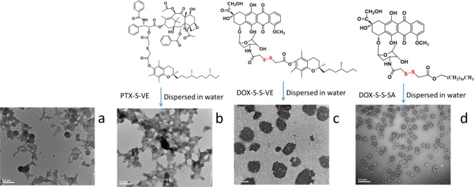
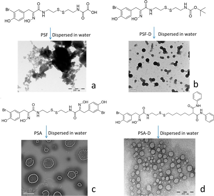
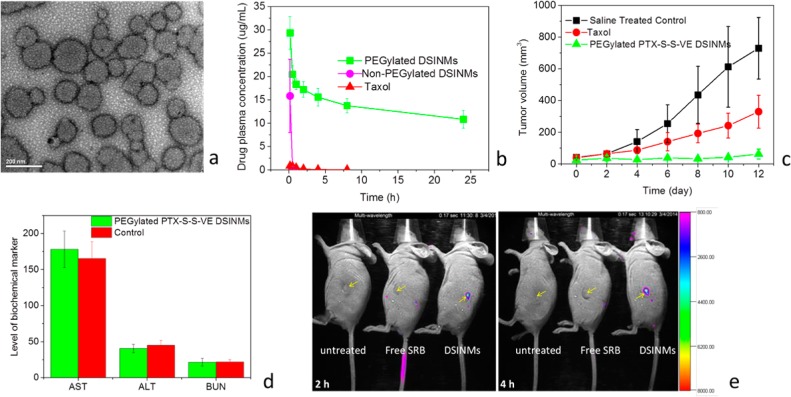
It is commonly observed that hydrophobic molecules alone cannot self-assemble into stable nanoparticles, requiring amphiphilic or ionic materials to support nanoparticle stability and function in vivo. We report herein newly self-assembled nanomedicines through entirely different mechanisms. We present proof-of-concept methodology and results in support of our hypothesis that disulfide-induced nanomedicines (DSINMs) are promoted and stabilized by the insertion of a single disulfide bond into hydrophobic molecules, in order to balance the competition between intermolecular forces involved in the self-assembly of nanomedicines. This hypothesis has been explored through diverse synthetic compounds, which include four first-line chemotherapy drugs (paclitaxel, doxorubicin, fluorouracil, and gemcitabine), two small-molecule natural products and their derivatives, as well as a fluorescent probe. Such an unprecedented and highly reproducible system has the potential to serve as a synthetic platform for a wide array of safe and effective therapeutic and diagnostic nanomedicine strategies.

Keywords: disulfide bond; nanomaterials; nanomedicines; nanoparticles; prodrug; self-assemble.


Figures

Figure 1
Figure 1
Characterization of various hydrophobic drugs dispersed in water. (a–c) Molecular structures of PTX, VE and PTX–VE and TEM images after dispersion in water. The sample in (b) (VE) was stained on the TEM grid immediately after the self-assembling process. Molecular structure of PTX–S–S–VE and TEM (d) and SEM (d) image of PTX–S–S–VE DSINMs. (f) Particle size distribution of PTX–S–S–VE DSINMs in DI water stored at 4 °C for two months. Scale bar: (a–b), 1 μm; (c–d), 0.2 μm; (e), 100 nm.
Figure 2
Figure 2
Illustration of the role of disulfide bond in self-assembly of DSINMs. (a) TEM image of PTX–S–S–VE DSINMs after incubation in 2.5 mM GSH for 0.5 h. (b) Molecular structure of PTX–S–VE and TEM image of PTX–S–VE after dispersion in water. (c–d) Molecular structures of DOX–S–S–VE and DOX–S–S–SA and TEM images of DOX–S–S–VE and DOX–S–S–SA DSINMs. Scale bar: (a–c), 0.2 μm; (d), 0.5 μm.
Figure 3
Figure 3
Molecular structures and TEM images of natural histone deacetylase inhibitors (HDACIs) and their derivatives after dispersion in water. (a) PSF, (b) PSF–D, (c) PSA, (d) PSA–D. Scale bar: (a–b), 0.2 μm; (c), 1 μm; (d), 0.2 μm.
Figure 4
Figure 4
Characterization of the structure and formation of DSINMs. (a) MD simulations of tetrameric PTX–S–S–VE in water. The carbons are colored according to their molecule of origin: blue (VE), orange (PTX), and yellow (S–S). (b) Imaging the dynamics of crystal growth. Ethanol containing PTX–VE and PTX–S–S–VE was placed onto a glass slide. After drying and desiccating, a drop of water was added to each sample, the slides were placed into a humidified chamber at room temperature for various hydration times and pictures were taken. The left panel (I, III, and V) shows PTX–VE and the right panel (II, IV, and VI) PTX–S–S–VE. (c) The electrostatic potential map of PTX–S–S–VE.
Figure 5
Figure 5
Characterization of PEGylated DSINMs for their physical image, plasma concentrations, anticancer activity, toxicity and tumor imaging. (a) TEM image of PTX–S–S–VE DSINMs PEGylated with 15% (w/w) DSPE-PEG2000. (b) Plasma concentration profiles of PEGylated and non-PEGylated PTX–S–S–VE DSINMs compared with Taxol (n = 3). (c) Antitumor effects in mice models, ** p < 0.01 (Student’s t-test, paired, two sided), compared with Taxol group and saline group (n = 5). (d) Kidney and liver function parameters in PEGylated PTX–S–S–VE DSINM and saline treated control groups (n = 5). (e) Tumor imaging in live mice. The tumor (indicated by arrows) bearing mice were imaged 2 and 4 h after injection of free SRB and PEGylated SRB–S–S–VE DSINMs. Images at 8, 12, and 48 h are shown in Supporting Information Figure S9.

Comment in

References

    1. Shi J.; Votruba A. R.; Farokhzad O. C.; Langer R. Nano Lett. 2010, 10, 3223–3230. - PMC - PubMed
    1. Han W.; Lin Z. Angew. Chem., Int. Ed. 2012, 51, 1534–46. - PubMed
    1. Clancy P. Nat. Nanotechnol. 2011, 6, 540–1. - PubMed
    1. Bolton J.; Bailey T. S.; Rzayev J. Nano Lett. 2011, 11, 998–1001. - PubMed
    1. Ernsting M. J.; Tang W. L.; MacCallum N. W.; Li S. D. Biomaterials 2012, 33, 1445–54. - PubMed

Publication types

MeSH terms