Review
. 2014 Sep 9;64(10):1033-46.
doi: 10.1016/j.jacc.2014.07.014.

Coronary artery manifestations of fibromuscular dysplasia

Affiliations
Review

Coronary artery manifestations of fibromuscular dysplasia

Katherine C Michelis et al. J Am Coll Cardiol. .

Abstract

Fibromuscular dysplasia (FMD) involving the coronary arteries is an uncommon but important condition that can present as acute coronary syndrome, left ventricular dysfunction, or potentially sudden cardiac death. Although the classic angiographic "string of beads" that may be observed in renal artery FMD does not occur in coronary arteries, potential manifestations include spontaneous coronary artery dissection, distal tapering or long, smooth narrowing that may represent dissection, intramural hematoma, spasm, or tortuosity. Importantly, FMD must be identified in at least one other noncoronary arterial territory to attribute any coronary findings to FMD. Although there is limited evidence to guide treatment, many lesions heal spontaneously; thus, a conservative approach is generally preferred. The etiology is poorly understood, but there are ongoing efforts to better characterize FMD and define its genetic and molecular basis. This report reviews the clinical course of FMD involving the coronary arteries and provides guidance for diagnosis and treatment strategies.

Keywords: acute coronary syndrome; coronary vessel anomalies; fibromuscular dysplasia; left ventricular dysfunction; myocardial infarction.


Figures

FIGURE 1
FIGURE 1. Angiographic Classification of Noncoronary FMD: Multifocal Versus Focal
(A) Multifocal renal artery FMD with alternating stenosis and dilation, creating the classic “string of beads” appearance on angiography (arrows). (B) Angiography showing focal renal artery FMD (arrows). FMD = fibromuscular dysplasia.
FIGURE 2
FIGURE 2. SCAD of the LAD Artery
A 34-year-old female patient presented with ST-segment elevation myocardial infarction 7 days after cesarean delivery of a healthy newborn. The pregnancy was complicated by hypertension. (A) Coronary angiography showing the left coronary system in a cranial projection, revealing extensive dissection of the mid-LAD coronary artery (arrows). (B) Optical coherence tomography of the dissected LAD artery, showing the true lumen (TL) and false lumen (FL). (C) Angiographically normal right coronary artery of the same patient. Original images are presented here, but this case was previously reported (124). LAD = left anterior descending; SCAD = spontaneous coronary artery dissection.
FIGURE 3
FIGURE 3. SCAD of the Left Circumflex Artery
A 37-year-old female patient presented with a troponin-positive non-ST-segment myocardial infarction. The patient had a pre-existing diagnosis of FMD with renal and carotid artery involvement. (A) Coronary angiography showing the left coronary system in a right anterior oblique-caudal projection. Arrows indicate dissection involving the obtuse marginal branch of the left circumflex with mottled appearance, narrowing, and filling defect. Note the normal, smooth appearance of the other vessels and segments. (B) Angiographically normal right coronary artery of the same patient. (C) Computed tomographic angiography of the carotid and vertebral arteries. Note the tortuosity and beading in the mid and distal portion of the right internal carotid artery (arrows), typical of multifocal FMD. The carotid bifurcation (the site where atherosclerosis occurs) is normal. Abbreviations as in Figures 1 and 2.
FIGURE 4
FIGURE 4. SCAD of the LAD Artery With Intramural Hematoma
A 40-year-old woman presented 10 days after a third-trimester miscarriage with troponin-positive non-ST-segment elevation myocardial infarction. (A) Coronary angiography revealed SCAD of the mid-LAD artery (white arrows). No stent was placed, and she was managed medically with a heparin drip and tight blood pressure control for 48 h. She was then discharged from the hospital on aspirin, clopidogrel, a beta-blocker, and a statin. She returned 5 days post-discharge with recurrent non-ST-segment elevation myocardial infarction and underwent (B) repeat coronary angiography that showed worsening dissection of the LAD artery (white arrows). Due to ongoing cardiac ischemia and worsening dissection, the proximal and mid-LAD arteries were then stented (white arrows) (C). (D) Caudal coronary angiographic view after stenting, with re-expansion of the LAD artery but smooth narrowing of the mid-distal left main artery (red arrow), ostial LAD (yellow arrow), and ramus intermedius (white arrow). (E and F) Intravascular ultrasound performed immediately after percutaneous intervention revealed a large intramural hematoma (asterisks) in the mid-distal left main artery (red arrow), which extended into the LAD artery (yellow arrow). Over the next 2 days, she continued to have intermittent chest pain and rising troponin levels; she therefore underwent a third cardiac catheterization and was found to have (G) new severe proximal narrowing of the left circumflex artery (white arrows) and an occluded first obtuse marginal artery (black arrow), with patent intervention sites. No further intervention was performed, and the patient improved with medical therapy over the following days. Transthoracic echocardiography revealed a mild-moderately decreased ejection fraction (46%), and she was ultimately discharged home on aspirin, clopidogrel, a beta-blocker, and an angiotensin-converting enzyme inhibitor. She has remained well for 1 month post-discharge and awaits formal evaluation for FMD. Abbreviations as in Figures 1 and 2.
FIGURE 5
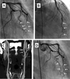
FIGURE 5. Smooth Coronary Artery Narrowing in a Patient With FMD
A 52-year-old woman with a history of migraine presented with non-ST-segment elevation myocardial infarction and was found to have smooth narrowing and distal tapering involving the LAD and left circumflex arteries. (A) Coronary angiography with caudal view showing distal smooth tapering of the LAD and left circumflex arteries (arrows). (B) Cranial view showing distal smooth tapering of the LAD artery (arrows). Several aliquots of both intracoronary nitroglycerin and intracoronary verapamil were administered over a 15-min period without any effect. (C) In the same patient, computed tomographic angiography of the carotid arteries is notable for a beaded appearance of the mid and distal portions of the cervical left internal carotid artery (arrows), consistent with FMD. The patient was managed conservatively with aspirin and clopidogrel. (D) Repeat coronary angiography 1 month after initial cardiac catheterization was angiographically normal, likely representing healed dissection of the LAD and left circumflex arteries (arrows). Abbreviations as in Figures 1 and 2.
FIGURE 6
FIGURE 6. Standing Waves
Arteriography of the axillary artery showing standing waves (arrows). Standing waves are due to catheter-induced spasm, and typically the administration of nitroglycerin will result in a normal-appearing artery. Note the regular oscillations that occur with standing waves, in contrast to the appearance of the “string of beads” (Figure 1), where the “beads” are larger than the normal caliber of the artery.
FIGURE 7
FIGURE 7. Myocardial Bridging
Coronary angiography showing cranial and caudal views, highlighting the “milking effect” that occurs with an LAD artery myocardial bridge, arising when the LAD artery takes an intramyocardial course. During systole, myocardial tissue surrounding the mid-LAD artery contracts, producing a narrowed segment of tunneled artery (arrows, lower panels). This narrowing is released during diastole (arrows, upper panels), when there is ventricular relaxation. Abbreviation as in Figure 2.
CENTRAL ILLUSTRATION
CENTRAL ILLUSTRATION. Angiographic Features of Fibromuscular Dysplasia Involving the Coronary Arteries
(A) Dissection, (B) smooth narrowing, (C) intramural hematoma, and (D) tortuosity. Spasm may also occur but is angiographically similar in appearance to smooth narrowing and is differentiated by intracoronary vasodilator administration.

References

    1. Slovut D, Olin J. Fibromuscular dysplasia. N Engl J Med. 2004;350:1862–1871. - PubMed
    1. Olin JW, Froehlich J, Gu X, et al. The United States Registry for Fibromuscular Dysplasia: results in the first 447 patients. Circulation. 2012;125:3182–3190. - PubMed
    1. Olin JW, Sealove BA. Diagnosis, management, and future developments of fibromuscular dysplasia. J Vase Surg. 2011;53:826–836. - PubMed
    1. Touze E, Oppenheim C, Trystram D, et al. Fibromuscular dysplasia of cervical and intracranial arteries. Int J Stroke. 2010;5:296–305. - PubMed
    1. Saw J, Ricci D, Starovoytov A, et al. Spontaneous coronary artery dissection: prevalence of predisposing conditions including fibromuscular dysplasia in a tertiary center cohort. J Am Coll Cardiol Intv. 2013;6:44–52. - PubMed

Publication types

MeSH terms