. 2014 Sep 5:2:4802.
doi: 10.1038/ncomms5802.

Somatic mutations in DROSHA and DICER1 impair microRNA biogenesis through distinct mechanisms in Wilms tumours

Affiliations

Somatic mutations in DROSHA and DICER1 impair microRNA biogenesis through distinct mechanisms in Wilms tumours

Dinesh Rakheja et al. Nat Commun. .

Erratum in

Abstract

Wilms tumour is the most common childhood kidney cancer. Here we report the whole-exome sequencing of 44 Wilms tumours, identifying missense mutations in the microRNA (miRNA)-processing enzymes DROSHA and DICER1, and novel mutations in MYCN, SMARCA4 and ARID1A. Examination of tumour miRNA expression, in vitro processing assays and genomic editing in human cells demonstrates that DICER1 and DROSHA mutations influence miRNA processing through distinct mechanisms. DICER1 RNase IIIB mutations preferentially impair processing of miRNAs deriving from the 5'-arm of pre-miRNA hairpins, while DROSHA RNase IIIB mutations globally inhibit miRNA biogenesis through a dominant-negative mechanism. Both DROSHA and DICER1 mutations impair expression of tumour-suppressing miRNAs, including the let-7 family, important regulators of MYCN, LIN28 and other Wilms tumour oncogenes. These results provide new insights into the mechanisms through which mutations in miRNA biogenesis components reprogramme miRNA expression in human cancer and suggest that these defects define a distinct subclass of Wilms tumours.


Conflict of interest statement

The authors declare no competing financial interests.

Figures

Figure 1
Figure 1. Mutations in the miRNA biogenesis pathway in Wilms tumours.
(a) Summary of somatic mutations detected by exome capture and massively parallel sequencing of matched Wilms tumours and germline DNA in the discovery set. (b) Mutations in miRNA biogenesis components are mutually exclusive with WT1 and CTNNB1 mutations. (c) Domain structure of DICER1 and DROSHA, indicating positions of mutations. Metal-binding residues in the RNase III domains are indicated in red. PAZ, Piwi–Argonaute–Zwille domain; DRBM, double-stranded RNA-binding motif. (d) Electropherograms from resequencing of selected mutations. Pie charts depict read counts from Illumina sequencing for reference (blue) and variant (red) alleles. (e) Modelling of G1809V mutation (silver) in the DICER1 metal-binding site, based on the structure of G. lamblia RNase III (PDB 2QVW). Numbering of equivalent Homo sapiens DICER1 residues is indicated. Dots indicate solvent-accessible surface.
Figure 2
Figure 2. Wilms tumour DICER1 and DROSHA mutations impair pri- and pre-miRNA processing in vitro.
(a) Schematic of pri-miRNA indicating RNase III cleavage sites. (b) DICER1 mutations affect pre-miRNA processing. Wild-type or mutant DICER1 was purified by immunoprecipitation and tested against pre-miRNA substrates. Arrowhead, mature miRNA; asterisk, processing intermediate formed by DICER1 RIIIA cleavage in the absence of RIIIB activity. (c) Mutations in the RNase III domains impair DROSHA activity. Wild-type or mutant DROSHA was purified by immunoprecipitation and tested against pri-miRNA substrates. Arrowhead, processed pre-miRNA.
Figure 3
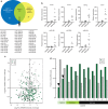
Figure 3. DICER1 and DROSHA mutations impair miRNA biogenesis in Wilms tumours.
(a) Summary of differentially expressed miRNAs in DICER1 and DROSHA-mutant tumours as determined by small RNA-seq. The 39 miRNAs whose expression is lower in both DICER1 and DROSHA-mutant tumours are listed. (b) Quantitative reverse transcription–PCR validation of expression of selected miRNAs in tumours with or without mutations in DROSHA and DICER1. (c) Fold-change in expression of 5′ (green) and 3′ (grey)-derived miRNAs comparing DROSHA or DICER1 mutant tumours with non-mutant tumours. Members of the let-7 family are indicated. (d) Fraction of total miRNA reads deriving from the 5′ (green) or 3′ (grey) arm of the pre-miRNA hairpin for each tumour. Arrowheads indicate lower representation of 5′ miRNAs in DICER1 mutants.
Figure 4
Figure 4. The heterozygous DROSHA E1147K mutation acts as a dominant-negative allele.
(a) TALEN-induced frameshift mutations in DROSHA+/− cells (left) and knock-in mutations in DROSHA+/E1147K cells (right) are shown. Silent mutations, incorporated to prevent re-cleavage by TALENs, are denoted by grey underlined letters, while the non-synonymous mutation is in red. Intronic sequences in lower case. (b) Immunoblotting of DROSHA levels in parental and mutant cell lines. The full blot is shown in Supplementary Fig. 4. (c) Distribution of miRNA expression levels, displayed as log2 (normalized expression), calculated by subtracting average ΔCt of each miRNA (CtmiRNA−CtU6, averaged within each genotype) from the median parental ΔCt value. Distributions compared by paired two-tailed t-test; *P<10−10. (d) Scatter plot of log2 (normalized expression) of individual miRNAs in DROSHA+/− and DROSHA+/E1147K cells versus parental cells. Red dots represent miRNAs whose expression differed significantly (P<0.05) between mutant cells and wild type; P-values computed by two-tailed t-test. (e) Expression of let-7 family members in DROSHA-mutant cells, relative to parental cells. Individual TaqMan assays for each miRNA were performed in triplicate for each of three clones within each genotype. Error bars represent mean±s.d. across three clones within each genotype. Dashed line represents expression in parental cells; P-values calculated by comparing expression in mutant versus parental cells by z-test. *P<0.05; **P<0.005.

References

    1. Chu A. et al.. Wilms’ tumour: a systematic review of risk factors and meta-analysis. Paediatr. Perinat. Epidemiol. 24, 449–469 (2010). - PubMed
    1. Rivera M. N. & Haber D. A. Wilms’ tumour: connecting tumorigenesis and organ development in the kidney. Nat. Rev. Cancer 5, 699–712 (2005). - PubMed
    1. Ko E. Y. & Ritchey M. L. Current management of Wilms’ tumor in children. J. Pediatr. Urol. 5, 56–65 (2009). - PubMed
    1. Kalapurakal J. A. et al.. Management of Wilms’ tumour: current practice and future goals. Lancet Oncol. 5, 37–46 (2004). - PubMed
    1. Murphy W. M., Grignon D. J. & Perlman E. J. inAFIP Atlas of Tumor Pathology Series 4. Tumors of the Kidney, Bladder, and Related Urinary Structures ed. Silverberg S. G. American Registry of Pathology (2004).

Associated data