. 2014 Nov 15;464(1):99-108.
doi: 10.1042/BJ20140420.

MEKK2 regulates paxillin ubiquitylation and localization in MDA-MB 231 breast cancer cells

Affiliations

MEKK2 regulates paxillin ubiquitylation and localization in MDA-MB 231 breast cancer cells

Magdalene Ameka et al. Biochem J. .

Abstract

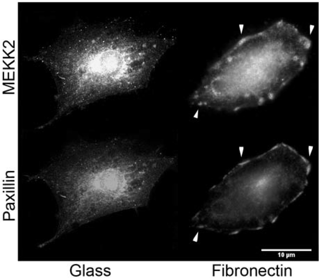
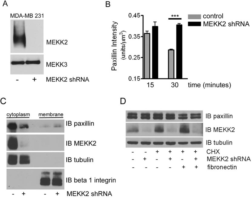
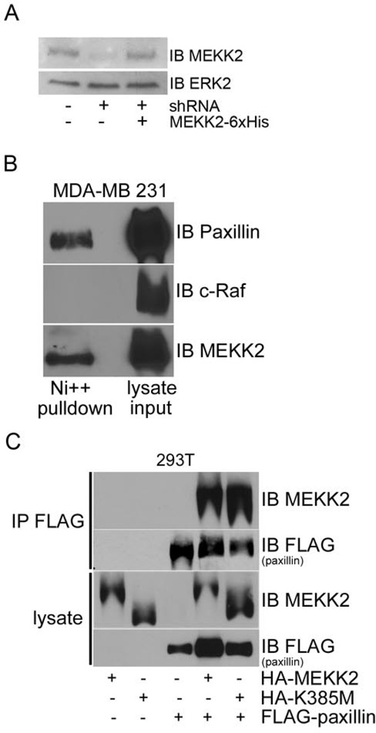
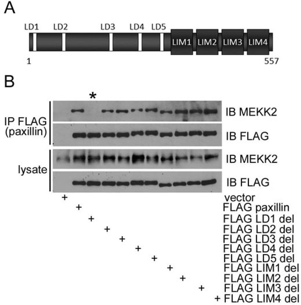
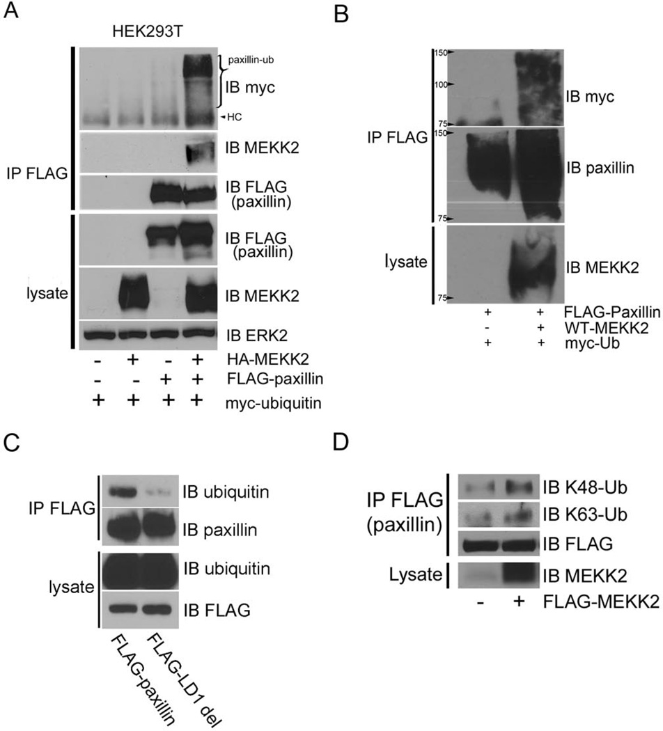
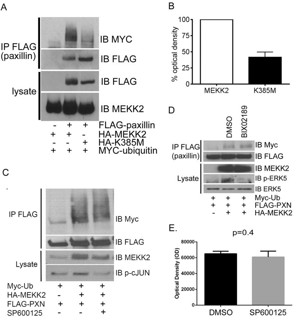
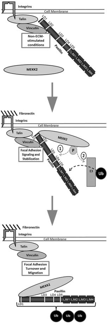
The intracellular kinase MEKK2 (mitogen-activated protein kinase/extracellular-signal-regulated kinase kinase kinase 2) is an upstream regulator of JNK (c-Jun N-terminal kinase), but additional functions for MEKK2 have not been well defined. Silencing MEKK2 expression in invasive breast tumour cells markedly inhibits xenograft metastasis, indicating that MEKK2 controls tumour cell function required for tumour progression. In our previous investigation of MEKK2 function, we discovered that tumour cell attachment to fibronectin recruits MEKK2 to focal adhesion complexes, and that MEKK2 knockdown is associated with stabilized focal adhesions and significant inhibition of tumour cell migration. In the present study we investigate MEKK2 function in focal adhesions and we report that MEKK2 physically associates with the LD1 motif of the focal adhesion protein paxillin. We reveal that MEKK2 induces paxillin ubiquitylation, and that this function requires both the paxillin LD1 motif and MEKK2 kinase activity. Finally, we demonstrate that MEKK2 promotes paxillin redistribution from focal adhesions into the cytoplasm, but does not promote paxillin degradation. Taken together, our results reveal a novel function for MEKK2 as a regulator of ubiquitylation-dependent paxillin redistribution in breast tumour cells.


Figures

Figure 1
Figure 1. MEKK2 co-localizes with paxillin in MDA-MB 231 cells adhered to fibronectin
(A) MDA-MB 231 breast tumor cells seeded on either uncoated glass coverslips (left) or on fibronectin-coated coverslips (right) for 16 hours, fixed and stained for immunofluorescence analysis with anti-MEKK2 or anti-paxillin antibodies. Areas of MEKK2 co-localization with paxillin are indicated by arrowheads. Scale bar represents 10 µm.
Figure 2
Figure 2. MEKK2 knockdown stabilizes paxillin localization in focal adhesions
(A) Anti-MEKK2 immunoblot of lysates from MDA-MB 231 cells that stably express either MEKK2 shRNA or empty vector control, showing specific MEKK2 knockdown (upper panel)subsequently the membrane was stripped and re-probed with anti-MEKK3 antibodies to show equal loading and knockdown specificity (lower panel). (B) Fluorescence microscopy with anti-vinculin and anti-paxillin antibodies was used to define paxillin localization in focal adhesions of MDA-MB 231 breast tumor cells attached to fibronectin. Graph depicting IMARIS quantification of paxillin co-localization with vinculin in three-dimensional focal adhesions over time. Comparison of the paxillin localized to focal adhesions in MDA-MB 231 cells (control) and cells with stable MEKK2 knockdown (MEKK2 shRNA) is shown. (C) Representative immunoblots showing the paxillin distribution into the cytoplasmic and membrane fractions from MDA-MB 231 cells expressing either MEKK2 shRNA or an empty shRNA vector control. Anti-tubulin and anti-beta 1 integrin antibody immunoblots were performed to confirm the purity of the cytoplasmic and membrane fractions, respectively. (D) Cells in suspension were exposed to cyclohexamide (40 µM) for one hour prior to attachment to fibronectin-coated plates for 30 minutes and then lysed and subjected to SDS-PAGE. Anti-paxillin immunoblot (upper panel) shows endogenous paxillin levels, whereas anti-MEKK2 blot shows knockdown efficiency (middle panel) and anti-tubulin blot (bottom panel) shows equal loading of protein. ***p<0.001. The data represented in the graphs was derived from at least three independent experiments.
Figure 3
Figure 3. Paxillin associates with MEKK2 independent of kinase activity
(A) Anti-MEKK2 immunoblot of MDA-MB 231 cells (left lane)with stable MEKK2 knockdown (center lane)and expression of “added back” shRNA-resistant 6xHis-tagged MEKK2 (right lane). (B) Immunoblot analysis shows the association between MEKK2 and paxillin in MDA-MB 231 breast tumor cells. Endogenous paxillin was co-purified with MEKK2 from MDA-MB 231 cells with stable expression of 6xHis-tagged MEKK2 (top panel) by Ni-NTA bead affinity column pull down. Also shown are immunoblots detecting c-Raf (middle panel, negative control) and MEKK2 (bottom panel). (C) HA-tagged wild type MEKK2 or kinase inactive mutant (K385M) MEKK2 co-immunoprecipitates with FLAG-tagged paxillin in transfected 293T cells (upper two panels). Immunoblots of lysates show loading in the lower two panels. The results are representative of at least three independent experiments.
Figure 4
Figure 4. Paxillin LD motif 1 is required for association with MEKK2
Immunoblot analysis shows MEKK2 co-immunoprecipitation with both wild type paxillin and mutant paxillin. (A) A representation of paxillin protein emphasizing the location of the LD motifs and LIM domains. (B) Eleven MDA-MB 231 cell lines were developed with stable expression of empty vector, FLAG-tagged wild type paxillin, or mutant paxillin. One interaction motif/domain was deleted from each paxillin mutant. Paxillin from each cell line was immunoprecipitated with anti-FLAG antibodies and the associated MEKK2 was detected by anti-MEKK2 immunoblot. The lane containing lysate from FLAG LD1 deletion mutant paxillin is indicated by the asterisk (*). The results are representative of at least three independent experiments.
Figure 5
Figure 5. MEKK2-induces paxillin ubiquitylation
(A) FLAG-paxillin, HA-MEKK2 and Myc-ubiquitin were co-expressed in 293T cells, and ubiquitylation of immunoprecipitated paxillin is revealed by anti-Myc immunoblot (top panel). Co-immunoprecipitated MEKK2 and total immunoprecipitated paxillin are shown in the second and third panels respectively. HC = Heavy Chain. (B) 293T cells transfected with FLAG-paxillin and HA-MEKK2 were lysed with 2% SDS and boiled to dissociate non-covalent protein interactions. FLAG-paxillin was then immunoprecipitated, and paxillin ubiquitylation was detected by anti-Myc immunoblot (upper panel)whereas the total immunoprecipitated paxillin is shown in the middle panel. MEKK2 expression was confirmed by lysate anti-MEKK2 immunoblot (bottom panel). (C) MDA-MB 231 cells that stably express FLAG-tagged wild type paxillin or LD1-deleted mutant paxillin were lysed and FLAG-tagged wild type or mutant paxillin was immunoprecipitated with anti-FLAG antibodies and separated by SDS-PAGE. Paxillin ubiquitylation was assessed by anti-ubiquitin immunoblot (upper panel), and immunoprecipitation efficiency was confirmed by anti-paxillin immunoblot (second panel). The expression of the tagged paxillin proteins was confirmed by anti-FLAG immunoblot of cell lysates (bottom panel), as was the ability of the anti-ubiquitin antibodies to detect endogenous ubiquitin (third panel). (D) Immunoblots with ubiquitin antibodies that specifically detect anti-K48-linked (upper panel) and anti-K63-linked ubiquitin (second panel) showing linkage of MEKK2-induced ubiquitylation of paxillin immunoprecipitated from transfected cell lysates. The membrane was re-probed with anti-FLAG antibodies (third panel) to demonstrate the effectiveness of the immunoprecipitation, and the bottom panel shows an anti-MEKK2 immunoblot of the cell lysates to confirm MEKK2 expression resulting from the transfected plasmid. The blots are representative of at least three independent experiments.
Figure 6
Figure 6. MEKK2 kinase activity-dependent promotion of paxillin ubiquitylation is independent of JNK or ERK5 activity
(A) FLAG-paxillin, HA-MEKK2 (wild type or inactive mutant K385M) and Myc-ubiquitin were co-expressed in 293T cells, and ubiquitylation of immunoprecipitated paxillin is revealed by anti-Myc immunoblot (top panel)and total immunoprecipitated paxillin is detected by anti-FLAG immunoblot (second panel). The expression levels of paxillin and MEKK2 in lysates were detected by anti-FLAG (third panel) and anti-MEKK2 (fourth panel) immunoblots, respectively. (B) Relative paxillin ubiquitylation is depicted in the bar graph showing densitometry analysis of anti-myc immunoblots from three independent experiments described in the Experimental section (A). (C and D) 293T cells transfected with FLAG-tagged paxillin, HA-tagged MEKK2, and myc-tagged ubiquitin expression vectors and treated with JNK inhibitor SP600125 (25 µM) (C) or MEK5 inhibitor BIX02189 (10 µM) (D) for 5 hours prior to lysis. Paxillin was immunoprecipitated with anti-FLAG antibodies and separated by SDS-PAGE. Paxillin ubiquitylation was assessed by anti-myc immunoblot. Inhibitor effectiveness was confirmed by phospho-specific immunoblots of JNK substrate c-Jun (C, bottom panel) and MEK5 substrate ERK5 (D, fourth panel) respectively. (E) Graph depicts comparison of optical density of ubiquitylated paxillin bands in either cells treated with JNK inhibitor SP600125 or vehicle (DMSO) alone as assessed with Image J software. Student’s t test analysis of in densitometry compiled from three independent experiments show p=0.4.
Figure 7
Figure 7. Model depicting MEKK2-dependent paxillin ubiquitylation and localization in invasive breast cancer cells
Cell attachment to fibronectin recruits active MEKK2 to focal adhesion complexes where it associates with paxillin. MEKK2 activity promotes paxillin ubiquitylation and subsequent re-distribution to the cytoplasm. Possible mechanisms by which MEKK2 activity-dependent ubiquitylation include 1) direct phosphorylation by MEKK2 targets paxillin for ubiquitylation, or 2) MEKK2 activates the E3 ligase that subsequently mediates paxillin ubiquitylation.

References

    1. Locascio A, Nieto MA. Cell movements during vertebrate development: integrated tissue behaviour versus individual cell migration. Curr. Opin. Genet. Dev. 2001;11:464–469. - PubMed
    1. Luster AD, Alon R, von Andrian UH. Immune cell migration in inflammation: present and future therapeutic targets. Nat. Immunol. 2005;6:1182–1190. - PubMed
    1. Machesky LM. Lamellipodia and filopodia in metastasis and invasion. FEBS. Lett. 2008;582:2102–2111. - PubMed
    1. Mackay CR. Moving targets: cell migration inhibitors as new anti-inflammatory therapies. Nat. Immunol. 2008;9:988–998. - PubMed
    1. Huttenlocher A, Horwitz AR. Integrins in cell migration. Cold Spring Harb. Perspect. Biol. 2011;3:a005074. - PMC - PubMed

Publication types