. 2014 Aug 13:2:96.
doi: 10.1186/s40478-014-0096-8.

Sequestration of latent TGF-β binding protein 1 into CADASIL-related Notch3-ECD deposits

Sequestration of latent TGF-β binding protein 1 into CADASIL-related Notch3-ECD deposits

Jessica Kast et al. Acta Neuropathol Commun. .

Abstract

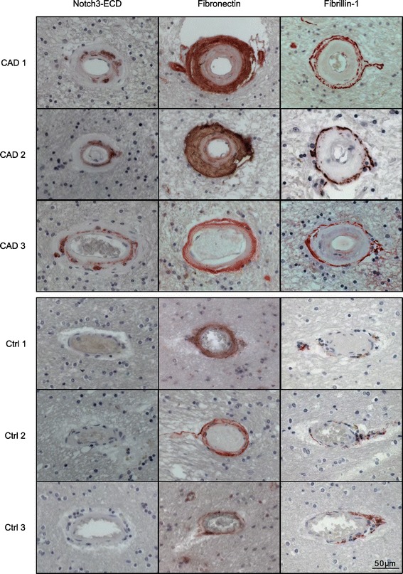
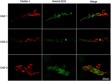
Introduction: Cerebral autosomal dominant arteriopathy with subcortical infarcts and leukoencephalopathy (CADASIL) represents the most common hereditary form of cerebral small vessel disease characterized by early-onset stroke and premature dementia. It is caused by mutations in the transmembrane receptor Notch3, which promote the aggregation and accumulation of the Notch3 extracellular domain (Notch3-ECD) within blood vessel walls. This process is believed to mediate the abnormal recruitment and dysregulation of additional factors including extracellular matrix (ECM) proteins resulting in brain vessel dysfunction. Based on recent evidence indicating a role for the transforming growth factor-β (TGF-β) pathway in sporadic and familial small vessel disease we studied fibronectin, fibrillin-1 and latent TGF-β binding protein 1 (LTBP-1), three ECM constituents involved in the regulation of TGF-β bioavailability, in post-mortem brain tissue from CADASIL patients and control subjects.

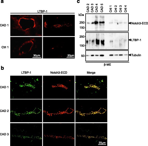
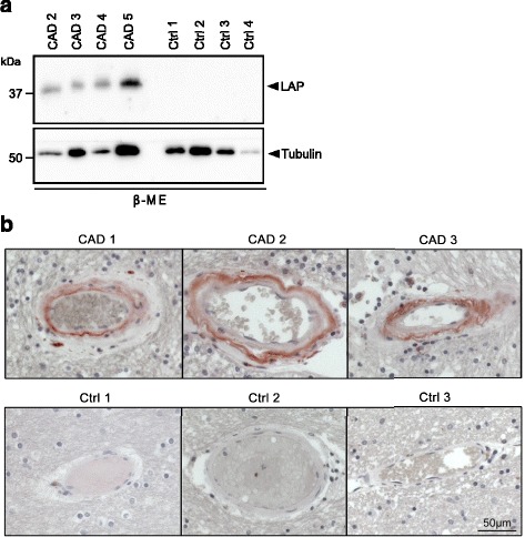
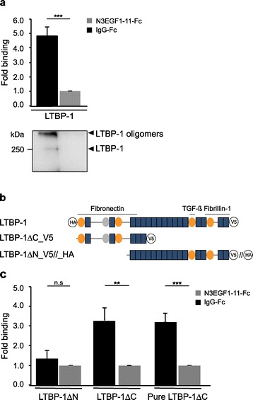
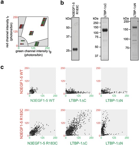
Results: Fibronectin and fibrillin-1 were found to be enriched in CADASIL vessels without co-localizing with Notch3-ECD deposits, likely as a result of fibrotic processes secondary to aggregate formation. In contrast, LTBP-1 showed both an accumulation and a striking co-localization with Notch3-ECD deposits suggesting specific recruitment into aggregates. We also detected increased levels of the TGF-β prodomain (also known as latency-associated peptide, LAP) indicating dysregulation of the TGF-β pathway in CADASIL development. In vitro analyses revealed a direct interaction between LTBP-1 and Notch3-ECD and demonstrated a specific co-aggregation of LTBP-1 with mutant Notch3.

Conclusion: We propose LTBP-1 as a novel component of Notch3-ECD deposits and suggest its involvement in pathological processes triggered by Notch3-ECD aggregation.


Figures

Figure 1
Figure 1
Fibronectin and fibrillin-1 accumulate in CADASIL brain vessels. Paraffin-embedded brain sections of three CADASIL patients and three healthy controls were stained for Notch3-ECD, fibronectin and fibrillin-1, counterstained with hematoxylin (blue) and analyzed by bright-field microscopy. (CAD: CADASIL patient; Ctrl: control).
Figure 2
Figure 2
Fibrillin-1 and Notch3-ECD do not co-localize. Frozen brain sections from three CADASIL patients were co-stained for fibrillin-1 and Notch3-ECD and analyzed by fluorescence microscopy. (CAD: CADASIL patient).
Figure 3
Figure 3
LTBP-1 accumulates in CADASIL brain vessels and co-localizes with Notch3-ECD aggregates. a Frozen brain sections from a CADASIL patient and a healthy control containing an arteriole (left panels) or a capillary (right panels) were stained for LTBP-1 and analyzed by fluorescence microscopy. b Frozen brain sections of three CADASIL patients were stained for LTBP-1 and Notch3-ECD and analyzed by confocal fluorescence microscopy. c LTBP-1 co-fractionates with Notch3-ECD. The final (β-ME-containing) fractions of sequentially extracted brain vessels from CADASIL patients and controls were immunoblotted for Notch3-ECD, LTBP-1 and β-tubulin. Shown is a representative blot from two independent experiments. (CAD: CADASIL patient; Ctrl: control).
Figure 4
Figure 4
LAP accumulates in CADASIL brain vessels. a LAP co-fractionates with Notch3-ECD. The β-ME fractions of sequentially extracted brain vessels from CADASIL patients and controls were immunoblotted for LAP and β-tubulin. Shown is a representative image of two independent extractions. b LAP accumulates in the tuncia media of CADASIL patient vessels. Paraffin-embedded brain sections were stained for LAP and counterstained with hematoxylin. (CAD: CADASIL patient; Ctrl: control).
Figure 5
Figure 5
LTBP-1 binds to immobilized Notch3 in a solid-phase binding assay. a The interaction of full-length LTBP-1 derived from conditioned cell supernatants with an immobilized Notch3 fragment (N3EGF1-11-Fc) is increased 4.9-fold when compared to a control ligand (IgG-Fc). Results are expressed as mean + SEM of seven independent experiments. Bound LTBP-1 was re-evaluated by immunoblotting and predominantly detected in oligomeric form. b Schematic representation of the used LTBP-1 constructs and their domain organization including cysteine-rich repeats (orange ovals), a hybrid domain (grey ovals), EGF-like repeats (blue boxes) and V5-His or HA tags (black circles). The fibronectin, TGF-β, and fibrillin-1 binding regions are indicated. Note that LTBP-ΔC-HA was used in assays with cell supernatants and LTBP-ΔC-V5 in assays requiring purified LTBP-1. c LTBP-1 binding to Notch3 is mediated by its N-terminus. While the N-terminal deletion variant LTBP-1ΔN does not bind significantly, the C-terminal deletion variant LTBP-1ΔC, either from conditioned supernatant or in purified form, shows significant interaction. Results are expressed as mean + SEM of five (LTBP-1ΔC_V5) and four (LTBP-1ΔN_HA) independent experiments. n.s.: not significant, ***p < 0.001; **p < 0.01; Mann–Whitney Test.
Figure 6
Figure 6
LTBP-1ΔC specifically co-aggregates with mutant Notch3. a Notch3-ECD multimer formation is monitored by SIFT and data illustrated in a 2D histogram: axes represent the intensity of photons per bin in the detector channel (green channel along the x-axis, red channel along the y-axis). While monomers and homomeric multimers result in data points in the lower left histogram area and along the axes respectively, heteromeric multimers are represented as high-intensity, dual-color signals in the white sector. b Purity of proteins used in the aggregation assay. Shown are silver-stained gels containing the elution fractions after metal-ion matrix (LTBP-1ΔC or LTBP-1ΔN) or Halo-tag-mediated purification (N3EGF1-5 R183C). c SIFT data from different protein combinations: while no high-molecular-weight particles are detected with N3EGF1-5 WT, typical aggregates are formed by N3EGF1-5 R183C and when combined with LTBP-1ΔC. In all other combinations homomeric multimers are detected indicating self-aggregation. Shown are representative images of 2–5 experiments. WT: wild-type.

References

    1. Pantoni L. Cerebral small vessel disease: from pathogenesis and clinical characteristics to therapeutic challenges. Lancet Neurol. 2010;9(7):689–701. doi: 10.1016/S1474-4422(10)70104-6. - DOI - PubMed
    1. Wardlaw JM, Smith C, Dichgans M. Mechanisms of sporadic cerebral small vessel disease: insights from neuroimaging. Lancet Neurol. 2013;12(5):483–497. doi: 10.1016/S1474-4422(13)70060-7. - DOI - PMC - PubMed
    1. Dichgans M. Genetics of ischaemic stroke. Lancet Neurol. 2007;6(2):149–161. doi: 10.1016/S1474-4422(07)70028-5. - DOI - PubMed
    1. Yamamoto Y, Craggs L, Baumann M, Kalimo H, Kalaria RN. Review: molecular genetics and pathology of hereditary small vessel diseases of the brain. Neuropathol Appl Neurobiol. 2011;37(1):94–113. doi: 10.1111/j.1365-2990.2010.01147.x. - DOI - PubMed
    1. Miao Q, Paloneva T, Tuominen S, Poyhonen M, Tuisku S, Viitanen M, Kalimo H. Fibrosis and stenosis of the long penetrating cerebral arteries: the cause of the white matter pathology in cerebral autosomal dominant arteriopathy with subcortical infarcts and leukoencephalopathy. Brain Pathol. 2004;14(4):358–364. doi: 10.1111/j.1750-3639.2004.tb00078.x. - DOI - PMC - PubMed

Publication types