

Axial eye growth and refractive error development can be modified by exposing the peripheral retina to relative myopic or hyperopic defocus
- PMID: 25190657
- PMCID: PMC4209715
- DOI: 10.1167/iovs.14-14524
Axial eye growth and refractive error development can be modified by exposing the peripheral retina to relative myopic or hyperopic defocus
Abstract
Purpose: Bifocal contact lenses were used to impose hyperopic and myopic defocus on the peripheral retina of marmosets. Eye growth and refractive state were compared with untreated animals and those treated with single-vision or multizone contact lenses from earlier studies.
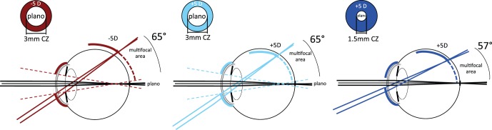
Methods: Thirty juvenile marmosets wore one of three experimental annular bifocal contact lens designs on their right eyes and a plano contact lens on the left eye as a control for 10 weeks from 70 days of age (10 marmosets/group). The experimental designs had plano center zones (1.5 or 3 mm) and +5 diopters [D] or -5 D in the periphery (referred to as +5 D/1.5 mm, +5 D/3 mm and -5 D/3 mm). We measured the central and peripheral mean spherical refractive error (MSE), vitreous chamber depth (VC), pupil diameter (PD), calculated eye growth, and myopia progression rates prior to and during treatment. The results were compared with age-matched untreated (N=25), single-vision positive (N=19), negative (N=16), and +5/-5 D multizone lens-reared marmosets (N=10).
Results: At the end of treatment, animals in the -5 D/3 mm group had larger (P<0.01) and more myopic eyes (P<0.05) than animals in the +5 D/1.5 mm group. There was a dose-dependent relationship between the peripheral treatment zone area and the treatment-induced changes in eye growth and refractive state. Pretreatment ocular growth rates and baseline peripheral refraction accounted for 40% of the induced refraction and axial growth rate changes.
Conclusions: Eye growth and refractive state can be manipulated by altering peripheral retinal defocus. Imposing peripheral hyperopic defocus produces axial myopia, whereas peripheral myopic defocus produces axial hyperopia. The effects are smaller than using single-vision contact lenses that impose full-field defocus, but support the use of bifocal or multifocal contact lenses as an effective treatment for myopia control.
Keywords: contact lenses; eye growth; myopia control; peripheral defocus; refractive error.
Copyright 2014 The Association for Research in Vision and Ophthalmology, Inc.
Figures


References
-
- Schaeffel F, Glasser A, Howland HC. Accommodation, refractive error and eye growth in chickens. Vision Res. 1988; 28: 639–657 - PubMed
-
- Norton TT, Siegwart JT. Animal models of emmetropization: matching axial length to the focal plane. J Am Optom Assoc. 1995; 66: 405–414 - PubMed
-
- Howlett MH, McFadden SA. Spectacle lens compensation in the pigmented guinea pig. Vision Res. 2009; 49: 219–227 - PubMed
-
- Graham B, Judge SJ. The effects of spectacle wear in infancy on eye growth and refractive error in the marmoset (Callithrix jacchus). Vision Res. 1999; 39: 189–206 - PubMed
Publication types
MeSH terms
Grants and funding
LinkOut - more resources
Full Text Sources
Other Literature Sources
Medical
