. 2015 Jun;11(3):511-25.
doi: 10.1007/s12015-014-9549-5.

Human vascular tissue models formed from human induced pluripotent stem cell derived endothelial cells

Affiliations

Human vascular tissue models formed from human induced pluripotent stem cell derived endothelial cells

David G Belair et al. Stem Cell Rev Rep. 2015 Jun.

Abstract

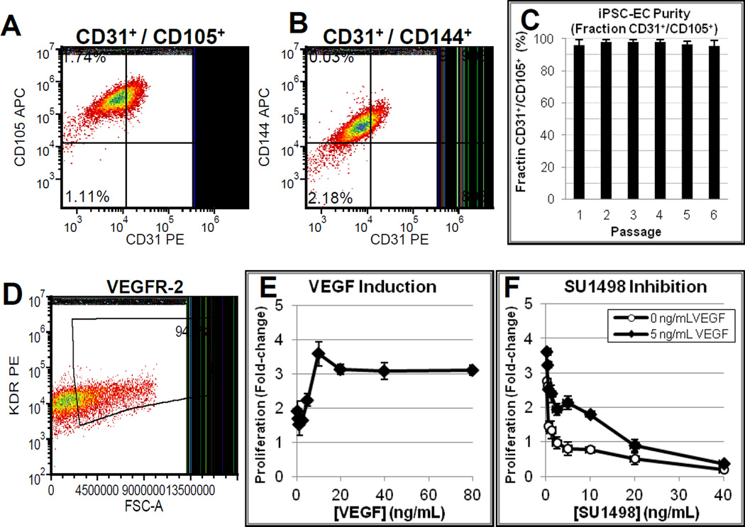
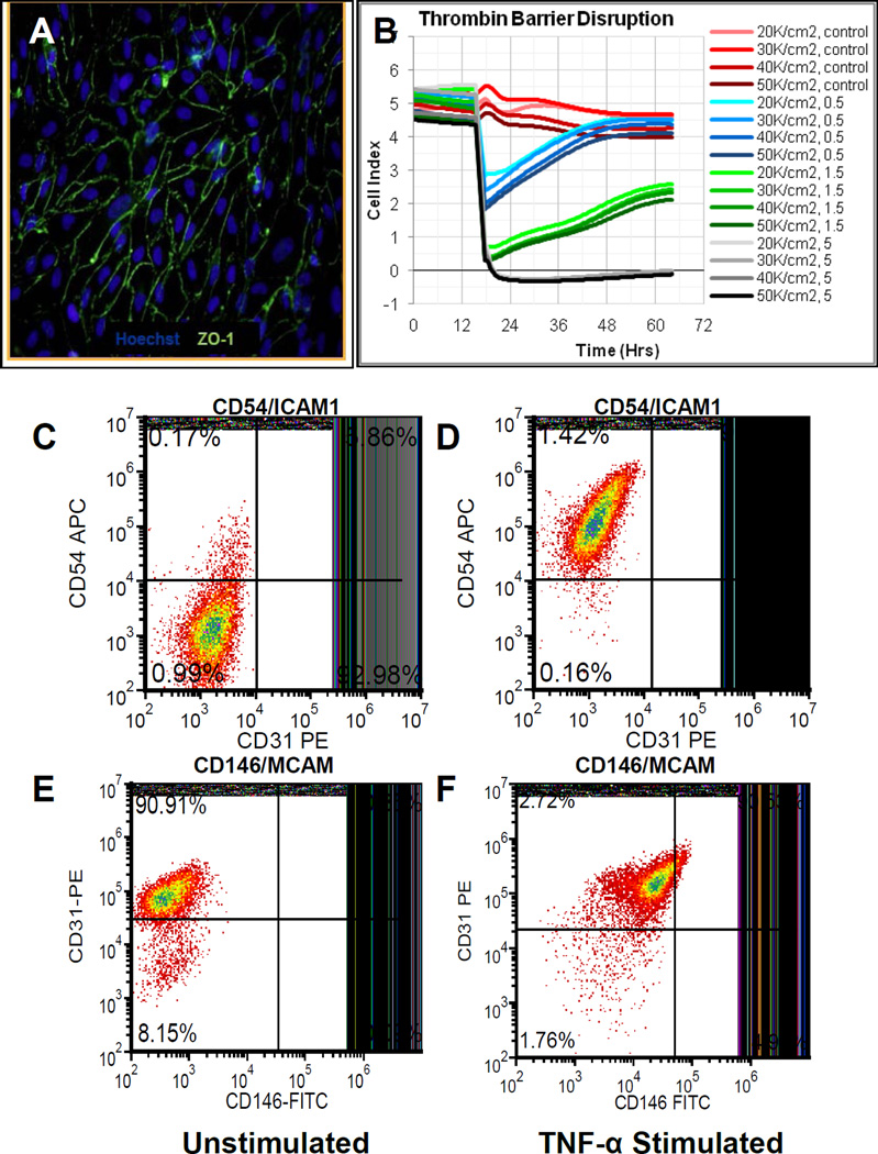
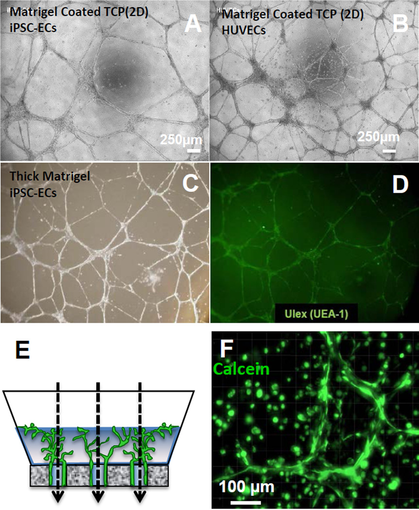
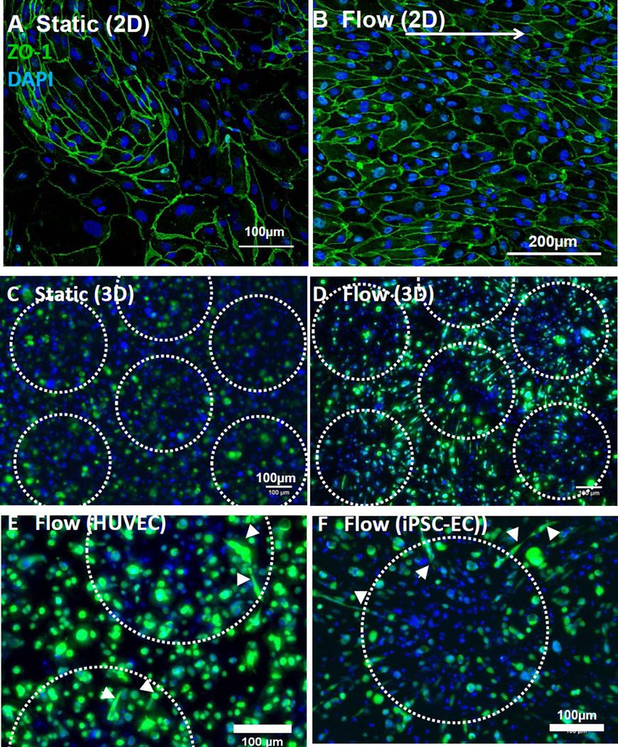
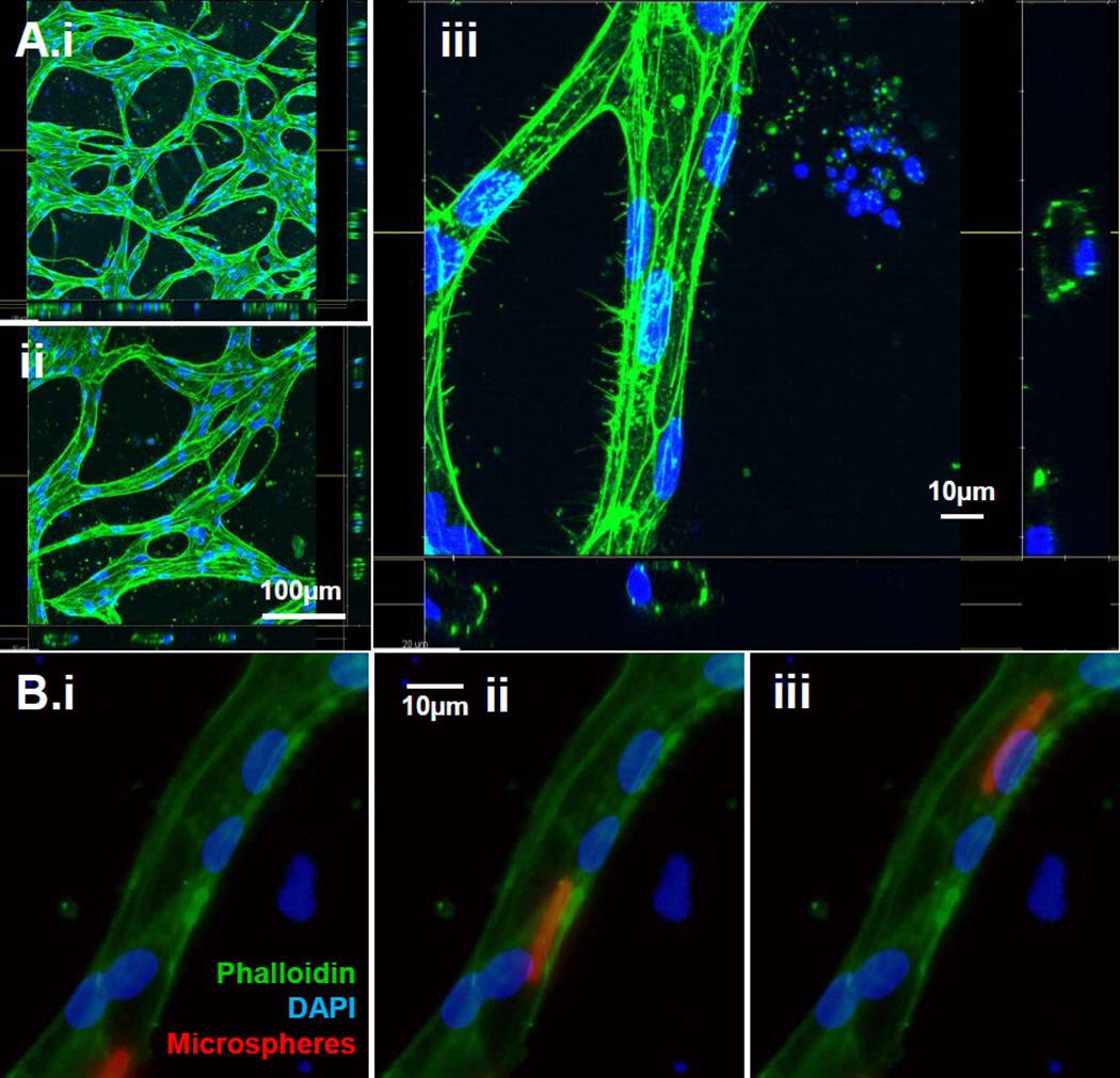
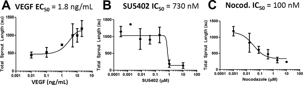
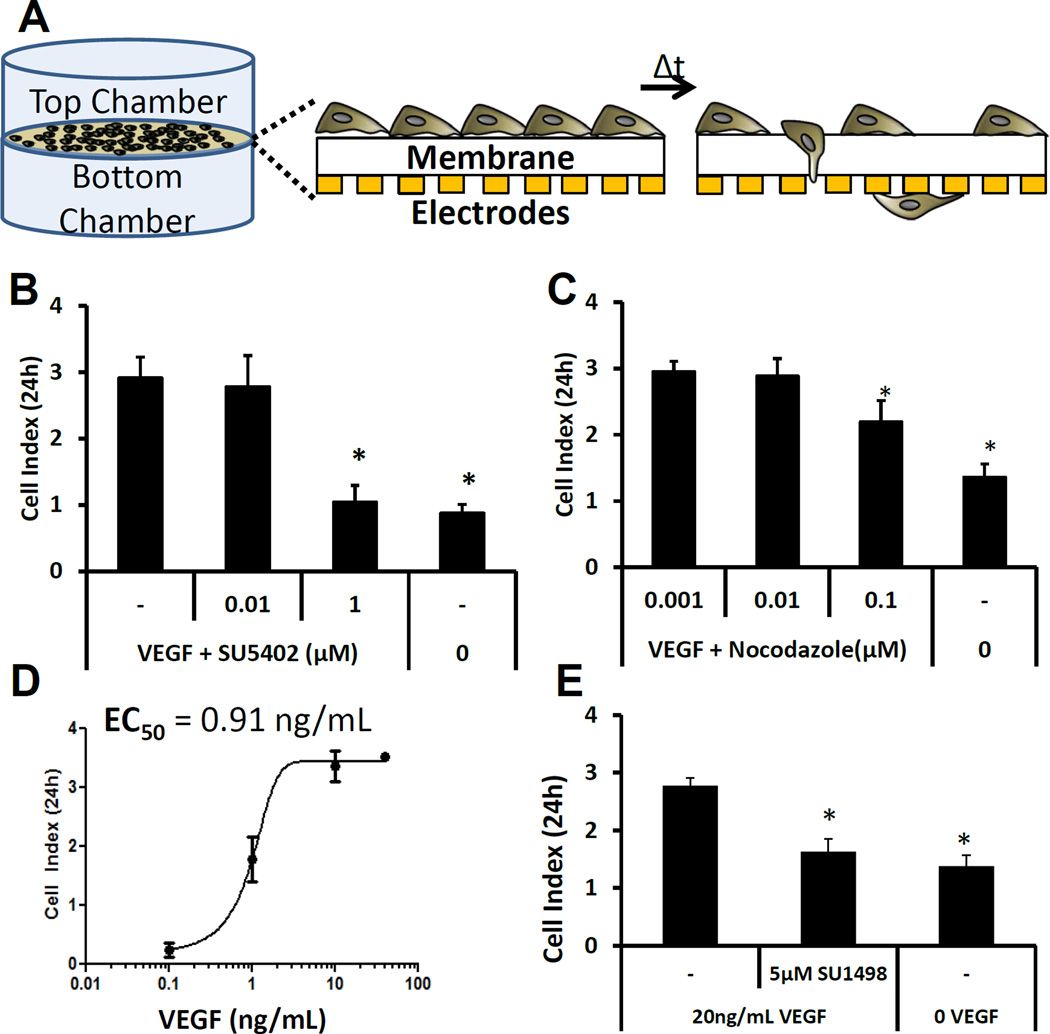
Here we describe a strategy to model blood vessel development using a well-defined induced pluripotent stem cell-derived endothelial cell type (iPSC-EC) cultured within engineered platforms that mimic the 3D microenvironment. The iPSC-ECs used here were first characterized by expression of endothelial markers and functional properties that included VEGF responsiveness, TNF-α-induced upregulation of cell adhesion molecules (MCAM/CD146; ICAM1/CD54), thrombin-dependent barrier function, shear stress-induced alignment, and 2D and 3D capillary-like network formation in Matrigel. The iPSC-ECs also formed 3D vascular networks in a variety of engineering contexts, yielded perfusable, interconnected lumen when co-cultured with primary human fibroblasts, and aligned with flow in microfluidics devices. iPSC-EC function during tubule network formation, barrier formation, and sprouting was consistent with that of primary ECs, and the results suggest a VEGF-independent mechanism for sprouting, which is relevant to therapeutic anti-angiogenesis strategies. Our combined results demonstrate the feasibility of using a well-defined, stable source of iPSC-ECs to model blood vessel formation within a variety of contexts using standard in vitro formats.


Conflict of interest statement

Conflict of Interest Disclosure: J.A.T. is a founder, stockowner, consultant, and board member of Cellular Dynamics International, Inc. D.A.M., R.L. and C.D. are employed by, and have a financial interest in, Cellular Dynamics International Inc.

Figures

Figure 1
Figure 1. iPSC-ECs stably express common endothelial cell markers and proliferate in response to VEGF/VEGFR2 signaling
(A) FACs analysis for PECAM / Endoglin co-expression. (B) FACs analysis illustrating PECAM / VE-cadherin co-expression. (C) iPSC-ECs maintain purity for at least 6 passages based on FACs analysis of CD31/CD105 co-expression. Average of 3 separate lots, 3 thaws each (9 samples total). Error bars = S.D. (D) FAC analysis for iPSC-EC expression of VEGFR-2 (KDR/Flk1). iPSC proliferation in response to (E) VEGF treatment (in Starvation Medium), and (F) when treated with the VEGFR-2 inhibitor SU1498 in Starvation Medium with 0 (white diamonds) or 5 ng/mL VEGF (black diamonds).
Figure 2
Figure 2. iPSC-EC expression and function
(A) iPSC-ECs express ZO-1 tight junction protein at cell borders. (B) iPSCs exhibit thrombin-dependent barrier function. Traces in red represent control conditions without thrombin. Blue, green, and black traces represent cell index over time in the presence of thrombin at 0.5 U/mL, 1.5 U/mL, or 5 U/mL respectively. (C–F) Upregulation of cell adhesion molecules (C–D) CD54 (ICAM1) and (E–F) CD146 (MCAM) for iPSC-ECs in response to treatment with 25 µg/mL TNF-α for 24 hours. ICAM1 expression (C) before and (D) after TNF-α treatment. MCAM expression (E) before and (F) after TNF-α treatment.
Figure 3
Figure 3. Tube forming assays for endothelial cells in 2D and 3D environments
(A–B) 2D tube-forming assay on Matrigel (coated TCP plate) for (A) iPSC-ECs and (B) HUVECs. (C–D) 3D tube-forming assay for iPSC-ECs in a thick layer of Matrigel; (C) Brightfield microscopy and (D) UEA-1 fluorescence imaging. UEA-1 is highly specific for EC lectin (fucose) [Jackson, JCS 1990]. (E) Schematic of 3D iPSC-EC encapsulation in Matrigel within a custom bioreactor to provide flow in the direction of the dashed arrows. (F) Maximum intensity projection of iPSC-ECs encapsulated in Matrigel (3D) and stained with Calcein-AM after 2 days of culture in the bioreactor from (E).
Figure 4
Figure 4. iPSC-EC organization when subjected to flow
(A–B) iPSC-ECs cultured in a microfluidics channel and subjected to lateral flow (shear stress = 20 dyn/cm2) to verify the capacity for alignment. Cells were randomly oriented without flow (A) but adopted elongated morphologies in the direction of an applied (left to right) shear stress (B). (C–D) Representative maximum intensity projection of iPSC-ECs encapsulated in Matrigel and cultured in a bioreactor for 4 days (as shown in Fig. 3E). Cells exhibited a rounded morphology in the absence of flow, but adopted an extended morphology towards the central axis of flow along the periphery of each channel. Channels were formed from 340µm pores (outlined in dotted white line) wherein flow was provided in the z-direction (into the frame of the micrographs. Z-stack = 300µm, z-slice thickness = 3µm (E–F) Representative maximum intensity projections of HUVECs (E) and iPSC-ECs (F) cultured in bioreactor (schematic Fig. 3E). Cells in the presence of flow exhibited an elongated morphology towards the central axis of flow (denoted by white arrowheads). Z-stack = 300µm, z-slice thickness = 3µm
Figure 5
Figure 5. iPSC-ECs form perfusable capillaries in a microfluidics device
iPSC-ECs encapsulated in fibrin within a central microfluidics channel with adjacent channels containing encapsulated fibroblasts. (A) After 4 days in culture with S1P-containing medium, iPSC-ECs encapsulated in central chamber were stained with FITC-Phalloidin and DAPI and were imaged using confocal laser scanning microscopy (A.i–ii). Scale bar: 100 µm (A.iii) Confocal image of iPSC-ECs stained with FITC-Phalloidin and DAPI, demonstrating that tubules contained a hollow lumen. Scale bar: 10µm (B) 4µm fluorescent microspheres were added to the channel and constrained to endothelial cell-lined tubes, demonstrating that iPSC-EC tubules were perfusable over time (Time scale = 119ms). Microspheres appear as a streak of ~15µm indicating movement in a given shutter exposure. Scale bar: 10 µm
Figure 6
Figure 6. VEGF/VEGFR2 signaling is necessary but not sufficient to induce iPSC-EC sprouting
(A) Schematic of cell spheroid formation in round-bottom low adhesion plates using Matrigel as a substrate to enable iPSC-EC sprouting. Analysis was performed after 7 total days in culture using Calcein-AM. (B) Quantification of total sprout length of iPSC-ECs cultured in a VEGF-only solution or Full Supplement (containing VEGF, FGF-2, IGF-1, and EGF) along with a library of pharmacological inhibitors. The control condition with 10 vol.% serum and no growth factors is shown by a dashed line, and +/1 standard deviation is shown as a gray bar. Statistical significance compared to the control is denoted for p-value < 0.05 (*). (C) Fluorescent micrographs demonstrating sprouting after 6 full days in culture with Matrigel. Cells were stained with Calcein-AM and Ethidium Homodimer-1. Scale bar represents 200 µm. Image series demonstrates sprouting in the presence of growth medium (C.i) with SU5402 (ii), nocodazole (iii.), or SU1498 (iv.). (D) Quantification of mean length of longest branched sprout in a subset of conditions in (B), showing increased longest continuous skeleton in SU1498-containing medium with Growth Medium. Statistical significance compared to the conditions in brackets is denoted for p-value < 0.05 (*). (E) Total sprout length as a function of VEGF in 2 and 10 vol.% serum with no inhibitor, SU1498, or Sunitinib. Statistical significance compared to no inhibitor conditions denoted for p-value < 0.05 (*)
Figure 7
Figure 7. iPSC-EC sprouting depends on both VEGF signaling and microtubule polymerization
(A) iPSC-EC total sprout length as a function of VEGF concentration. Sigmoidal regression analysis was used to calculate an EC50 value for VEGF. (B,C) Analysis of sprouting inhibition with SU5402 (B) and nocodazole (C) at a range of concentrations. Sigmoidal regression analysis was used to calculate an IC50 value for each inhibitor.
Figure 8
Figure 8. iPSC-EC migration is dependent on VEGF/VEGFR2 signaling and microtubule organization
(A,B) End-point analysis of iPSC-EC migration quantified using real-time impedance measurements in different concentrations of SU5402 (A) or nocodazole (B) in medium containing 10 vol.% serum with 20ng/mL VEGF in the bottom chamber and 10 vol.% serum and no VEGF in the top chamber. Statistical significance compared to the control without inhibitor is denoted is denoted for p-value < 0.05 (*). (C) End-point analysis of iPSC-EC migration in different concentrations of VEGF, added only to the bottom chamber. Sigmoidal regression analysis was used to calculate an EC50 value for VEGF (D) End-point analysis of iPSC-EC migration in the presence of the VEGF receptor tyrosine kinase inhibitor SU1498. Statistical significance compared to the no inhibitor control is denoted for p-value < 0.05 (*).

References

    1. Novosel EC, Kleinhans C, Kluger PJ. Vascularization is the key challenge in tissue engineering. Advanced Drug Delivery Reviews. 2011;63(4–5):300–311. - PubMed
    1. Phelps EA, García AJ. Engineering more than a cell: vascularization strategies in tissue engineering. Current Opinion in Biotechnology. 2010;21(5):704–709. - PMC - PubMed
    1. Nguyen EH, Zanotelli MR, Schwartz MP, Murphy WL. Differential effects of cell adhesion, modulus and VEGFR-2 inhibition on capillary network formation in synthetic hydrogel arrays. Biomaterials. 2014;35(7):2149–2161. - PMC - PubMed
    1. Moon JJ, Saik JE, Poche RA, Leslie-barbick JE, Smith AA, Dickinson ME, West JL. Biomimetic hydrogels with pro-angiogenic properties. Biomaterials. 2011;31(14):3840–3847. - PMC - PubMed
    1. Phelps EA, Landázuri N, Thulé PM, Taylor WR, García AJ. Bioartificial matrices for therapeutic vascularization. Proceedings of the National Academy of Sciences of the United States of America. 2010;107(8):3323–3328. - PMC - PubMed

Publication types

MeSH terms

Substances