. 2014 Sep 3;87(3):359-71.
eCollection 2014 Sep.

Arterial shear stress reduces eph-b4 expression in adult human veins

Affiliations

Arterial shear stress reduces eph-b4 expression in adult human veins

Lynn S Model et al. Yale J Biol Med. .

Abstract

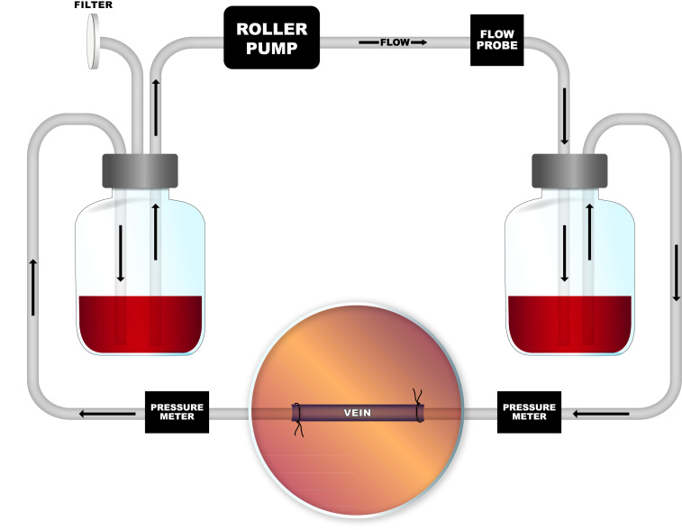
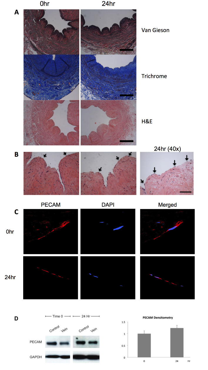
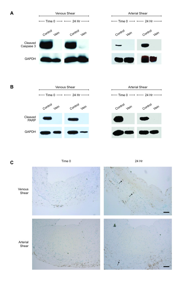
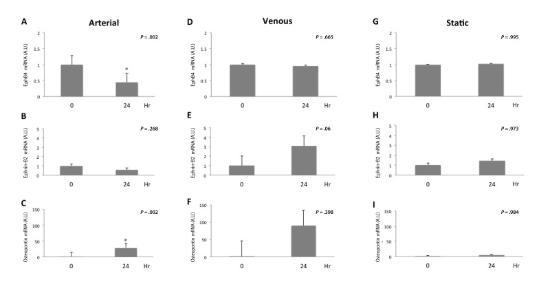
Vein graft adaptation to the arterial environment is characterized by loss of venous identity, with reduced Ephrin type-B receptor 4 (Eph-B4) expression but without increased Ephrin-B2 expression. We examined changes of vessel identity of human saphenous veins in a flow circuit in which shear stress could be precisely controlled. Medium circulated at arterial or venous magnitudes of laminar shear stress for 24 hours; histologic, protein, and RNA analyses of vein segments were performed. Vein endothelium remained viable and functional, with platelet endothelial cell adhesion molecule (PECAM)-expressing cells on the luminal surface. Venous Eph-B4 expression diminished (p = .002), Ephrin-B2 expression was not induced (p = .268), and expression of osteopontin (p = .002) was increased with exposure to arterial magnitudes of shear stress. Similar changes were not found in veins placed under venous flow or static conditions. These data show that human saphenous veins remain viable during ex vivo application of shear stress in a bioreactor, without loss of the venous endothelium. Arterial magnitudes of shear stress cause loss of venous identity without gain of arterial identity in human veins perfused ex vivo. Shear stress alone, without immunologic or hormonal influence, is capable of inducing changes in vessel identity and, specifically, loss of venous identity.

Keywords: EphB4; Ephrin-B2; Saphenous vein; bioreactor; osteopontin; shear stress; vein graft adaptation.


Figures

Figure 1
Figure 1
Bioreactor schematic. Digital programmable roller pump pushes fluid through system in a simple pulsatile fashion, set at desired rate for arterial or venous shear.
Figure 2
Figure 2
Vein structural and endothelial integrity after arterial shear stress. A. Photomicrographs of segments of adult human saphenous vein, 0 or 24 hours after shear stress treatment in the bioreactor. Staining with either Van Gieson, trichrome, or H&E stains. Scale bar represents 200 μm. B. Photomicrographs of saphenous vein segments, high power, stained with H&E. Arrows show endothelial cells on the luminal surface. Scale bar represents 50 μm. C. Immunofluourescence of the luminal surface of a vein segment, 0 or 24 hours of arterial shear, focused on individual endothelial cells. Red, PECAM; Blue, DAPI. N = 6, representative sample shown. D. Left panel, representative Western blot of PECAM and GAPDH in vein samples treated with 0 or 24 hours of arterial shear stress. Control Lane is HUVEC. N = 4. Right panel, bar graph shows mean densitometry of PECAM normalized to GAPDH, with time 0 as the reference; y-axis is in arbitrary units.
Figure 3
Figure 3
Vein viability after venous or arterial shear stress. A. Western blot for cleaved caspase-3 or GAPDH in representative vein samples at 0 or 24 hours of venous or arterial shear stress. Positive control; staurosporine-killed HUVEC. N = 6. B. Western blot for cleaved PARP or GAPDH in representative vein samples at 0 or 24 hours of venous and arterial shear stress. Positive control; staurosporine-killed HUVEC. N = 6. C. Photomicrographs showing representative TUNEL staining of matched vein samples at 0 or 24 hours of arterial (upper panels) or venous (lower panels) shear stress. Arrows indicate TUNEL positive cells. Scale bar represents 200 μm. N = 8.
Figure 4
Figure 4
Vein function and metabolic activity after arterial shear stress. A. Western blot and densitometry for mTOR or GAPDH in individual vein samples at 0 or 24 hours of arterial shear. Control, HUVECs. N = 8. B. Western blot and densitometry for eNOS or GAPDH in representative vein samples at 0 and 24 hours of arterial shear stress. Control, HUVECs. N = 8 C. Western blot and densitometry for phospho-eNOS or GAPDH in representative vein samples at 0 and 24 hours of arterial shear stress. Control, HUVECs. N = 8.
Figure 5
Figure 5
Eph-B4 expression is reduced with arterial but not venous shear stress treatment. A. Bar graph shows Eph-B4 mRNA transcript number, normalized to GAPDH, at 0 or 24 hours of arterial shear stress. n = 10; *, P = .002. B. Bar graph shows Ephrin-B2 mRNA transcript number, normalized to GAPDH, at 0 or 24 hours of arterial shear stress. n = 9; P = .268. C. Bar graph shows osteopontin mRNA transcript number, normalized to GAPDH, at 0 or 24 hours of arterial shear stress. n = 7; *, P =.002. D. Bar graph shows Eph-B4 mRNA transcript number, normalized to GAPDH, at 0 or 24 hours of venous shear stress. n = 4; P = .665. E. Bar graph shows Ephrin-B2 mRNA transcript number, normalized to GAPDH, at 0 or 24 hours of venous shear stress. n = 6; P = .06. F. Bar graph shows osteopontin mRNA transcript number, normalized to GAPDH, at 0 or 24 hours of venous shear stress. n = 4; P = .398. G. Bar graph shows Eph-B4 mRNA transcript number, normalized to GAPDH, at 0 or 24 hours of static conditions. n = 3, P = .995. H. Bar graph shows Ephrin-B2 mRNA transcript number, normalized to GAPDH, at 0 or 24 hours of static conditions. n = 3, P = .973. I. Bar graph shows osteopontin mRNA transcript number, normalized to GAPDH, at 0 or 24 hours of static conditions. n = 3, P = .984.
Figure 6
Figure 6
Eph-B4 immunofluorescence is reduced after 24 hours of arterial shear stress but not static conditions. Bar graphs demonstrate relative immunoreactivity quantification of arterial and venous conditions normalized to static conditions at both time 0 (P = .98) and 24 hours (P = .53).

References

    1. Bradbury AW, Adam DJ, Bell J, Forbes JF, Fowkes FGR, Gillespie I. et al. Bypass versus Angioplasty in Severe Ischaemia of the Leg (BASIL) trial: A survival prediction model to facilitate clinical decision making. J Vasc Surg. 2010;51(5):52S–68S. - PubMed
    1. Kocher O, Gabbiani G. Cytoskeletal features of normal and atheromatous human arterial smooth muscle cells. Hum Pathol. 1986;17(9):875–880. - PubMed
    1. Muto A, Model L, Ziegler K, Eghbalieh SDD, Dardik A. Mechanisms of Vein Graft Adaptation to the Arterial Circulation. Circ J. 2010;74(8):1501–1512. - PMC - PubMed
    1. Howard AB, Alexander RW, Nerem RM, Griendling KK, Taylor WR. Cyclic strain induces an oxidative stress in endothelial cells. Am J Physiol. 1997;272(2 Pt 1):C421–C427. - PubMed
    1. Schwartz LB, O’Donohoe MK, Purut CM, Mikat EM, Hagen PO, McCann RL. Myointimal thickening in experimental vein grafts is dependent on wall tension. J Vasc Surg. 1997;15(1):176–186. - PubMed

Publication types

Substances

LinkOut - more resources