Radiographic indicators of a shared epiphysis in radial polydactyly
- PMID: 25191163
- PMCID: PMC4152434
- DOI: 10.1007/s11552-013-9593-7
Radiographic indicators of a shared epiphysis in radial polydactyly
Abstract
Background: Thumb epiphyses cannot be visualized on radiographs in infants with radial polydactyly, making it difficult to classify by Wassel type. The purpose of this study was to identify radiographic features that distinguish a separate epiphysis from a shared epiphysis. This may assist in operative planning and establishing prognosis.
Methods: The charts of 34 radial polydactyly patients treated with surgical reconstruction from 2008 through 2012 were retrospectively reviewed. Measurements of the most proximal bones involved in the duplication, including length, width at shaft, width at base, distance between radial and ulnar thumb, and angle between radial and ulnar thumb, were taken from PA radiographs of the thumb by four blinded individuals. The interclass correlation coefficient was calculated to determine inter-observer reliability. Operative notes were reviewed to distinguish between shared and separate epiphyses. Several indices were created from these measurements.
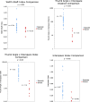
Results: Radiographic measurements showed high inter-observer reliability. There were statistically significant differences between patients with separate and shared epiphyses for indices for the width shaft index, interspace distance, the angle × interspace distance, and the angle × interspace index.
Conclusion: Radiographic differences exist between children with separate and shared epiphyses. In patients with shared epiphyses, the radial thumb tends to be smaller, closer to the ulnar thumb, and less divergent. Threshold values were identified for predicting the status of the epiphysis based on the angle × interspace distance and the angle × interspace index. These values may be used to help determine in advance of surgery if a shared epiphysis exists.
Keywords: Classification; Congenital; Duplication; Radiograph; Thumb; Wassel.
Figures


References
LinkOut - more resources
Full Text Sources
Other Literature Sources

