Review
. 2014 Aug 20:5:192.
doi: 10.3389/fphar.2014.00192. eCollection 2014.

The still mysterious roles of cysteine-containing glutathione transferases in plants

Affiliations
Review

The still mysterious roles of cysteine-containing glutathione transferases in plants

Pierre-Alexandre Lallement et al. Front Pharmacol. .

Abstract

Glutathione transferases (GSTs) represent a widespread multigenic enzyme family able to modify a broad range of molecules. These notably include secondary metabolites and exogenous substrates often referred to as xenobiotics, usually for their detoxification, subsequent transport or export. To achieve this, these enzymes can bind non-substrate ligands (ligandin function) and/or catalyze the conjugation of glutathione onto the targeted molecules, the latter activity being exhibited by GSTs having a serine or a tyrosine as catalytic residues. Besides, other GST members possess a catalytic cysteine residue, a substitution that radically changes enzyme properties. Instead of promoting GSH-conjugation reactions, cysteine-containing GSTs (Cys-GSTs) are able to perform deglutathionylation reactions similarly to glutaredoxins but the targets are usually different since glutaredoxin substrates are mostly oxidized proteins and Cys-GST substrates are metabolites. The Cys-GSTs are found in most organisms and form several classes. While Beta and Omega GSTs and chloride intracellular channel proteins (CLICs) are not found in plants, these organisms possess microsomal ProstaGlandin E-Synthase type 2, glutathionyl hydroquinone reductases, Lambda, Iota and Hemerythrin GSTs and dehydroascorbate reductases (DHARs); the four last classes being restricted to the green lineage. In plants, whereas the role of DHARs is clearly associated to the reduction of dehydroascorbate to ascorbate, the physiological roles of other Cys-GSTs remain largely unknown. In this context, a genomic and phylogenetic analysis of Cys-GSTs in photosynthetic organisms provides an updated classification that is discussed in the light of the recent literature about the functional and structural properties of Cys-GSTs. Considering the antioxidant potencies of phenolic compounds and more generally of secondary metabolites, the connection of GSTs with secondary metabolism may be interesting from a pharmacological perspective.

Keywords: cysteines; deglutathionylation; glutathione transferases; photosynthetic organisms; phylogeny.


Figures

Figure 1
Figure 1
Rooted phylogenetic tree of plant GSTs. The sequences used are those identified in Arabidopsis thaliana (Lan et al., 2009), Hordeum vulgare (Rezaei et al., 2013), Oryza sativa (Lan et al., 2009), Physcomitrella patens (Liu et al., 2013), Pinus tabulaeformis (Lan et al., 2013), Populus trichocarpa (Lan et al., 2009), and Solanum lycopersicum (Csiszar et al., 2014). Sequences were aligned with PROMALS3D and alignment manually adjusted with Seaview software (Gouy et al., 2010). The phylogenetic tree was constructed with BioNJ (Gascuel, 1997) in Seaview, rooted with E. coli glutaredoxin 2 and edited with Figtree software (http://tree.bio.ed.ac.uk/software/figtree/). The robustness of the branches was assessed by the bootstrap method with 500 replications. Various classes can be distinguished: Dehydroascorbate reductase (DHAR), Elongation factor 1Bγ (EF1Bγ), Glutathionyl hydroquinone reductase (GHR), Phi (GSTF), Hemerythrin (GSTH), Iota (GSTI), Lambda (GSTL), Theta (GSTT), Tau (GSTU), Zeta (GSTZ), Microsomal prostaglandin E synthase type 2 (mPGES-2), Tetrachloro-hydroquinone dehalogenase (TCHQD), and Ure2p. The scale marker represents 0.1 substitutions per residue. For clarity, the names of the sequences have not been indicated but all sequences are available in the Supplementary Material.
Figure 2
Figure 2
Unrooted phylogenetic tree of Cys-GSTs present in the green lineage. Sequences were aligned with PROMALS3D using 1Z9H, 3PPU, and 4PQH PDB structures as templates. Then the alignment has been manually adjusted with Seaview software. The phylogenetic tree was constructed with BioNJ and edited with Figtree software (http://tree.bio.ed.ac.uk/software/figtree/). The robustness of the branches was assessed by the bootstrap method with 500 replications. The scale marker represents 0.1 substitutions per residue. For clarity, the names of the sequences have not been indicated but all sequences are available in the Supplementary Material.
Figure 3
Figure 3
Amino acid alignment and protein architecture of plant Cys-GSTs. (A) Amino acid sequence alignment of three representative members from each Cys-GST class. The sequences were structurally aligned using PROMALS3D server using as references the solved structures of PtGSTL1 [PDB code 4PQH (Lallement et al., 2014)], PtGSTL3 [PDB code 4PQI (Lallement et al., 2014)], Phanerochaete chrysosporium GHR1 [PDB code 3PPU (Meux et al., 2011)], and Macaca fascicularis mPGES-2 [PDB code 1Z9H (Yamada et al., 2005)] since there is no structure available for DHARs, GSTIs, and GSTHs. Since the structure of poplar GSTL1 has been solved, its secondary structures have been indicated as reference using ESPript 3.0 (http://espript.ibcp.fr/ESPript/ESPript/index.php), with the helices and the arrows corresponding respectively to α-helices and to β-strands. Strictly conserved residues are marked in white characters on a red background, whereas residues with similar functional groups are in red characters on white background. The indicated numbering corresponds to that of PtGSTL1 which has been used as a whole. For clarity, N- and C-terminal extensions present in Cys-GSTs have been removed from the alignment to keep only the sequences corresponding to secondary structures forming the GST fold. At is for Arabidopsis thaliana, Pt for Populus trichocarpa, Os for Oryza sativa, Pp for Physcomitrella patens, Sm for Selaginella moellendorffii, and Vc for Volvox carteri. The catalytic cysteine (*), cis-proline (cis-Pro), residues stabilizing the γ-glutamate residue of glutathione (##) and N-cap residue are shown. The N-capping box is surrounded in green. (B) Schematic representation of the protein architecture of plant Cys-GSTs. The N-terminal Trx-like domain and the all-helical C-terminal domain are represented respectively in red and green. Blue boxes correspond to putative or confirmed targeting sequences. The orange box corresponds to the membrane anchoring tail of mPGES-2. Purple boxes represent N-terminal extensions that do not correspond to targeting sequences and gray boxes represent additional C-terminal domains. The position of the active site motif harboring the catalytic cysteine is indicated in black. The presence of inserted sequences in some classes corresponds to dashed lines in other classes. Secondary structures are shown as α-helices and β-strands. The size of the boxes is proportional to the length in amino acids.
Figure 4
Figure 4
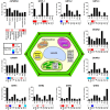
Subcellular localization and gene expression profiles of the different Arabidopsis thaliana Cys-GSTs. For each GST, the relative transcript expression is shown in relation to both plant developmental stages (bar-graphs) and perturbations (heatmaps). For clarity, the number of developmental conditions was reduced to eight classes, where each gene was normalized to its maximum expression within the selected dataset (see Materials and Methods). For the response to perturbations, the expression values were organized into five color-coded groups based on their log2-ratios. For clarity, the numerous perturbations included in the dataset were grouped into 11 classes: Biotic stress (Bs), Chemicals (Che), Germination (Ge), Light (Li), N-starvation (-N), Fe-deficiency (-Fe), Salt stress (Sa), Hypoxia (Hy), Drought (Dr), Heat (He), and Cold (Co).
Figure 5
Figure 5
Structural organization of Cys-GSTs. All structures are shown as cartoon with the N- and C-terminal domains colored in cyan and in purple, respectively. Glutathione (GSH) or glutathione adducts (GS) are represented as sticks. In F, glutathione is only present in monomer A. All figures have been prepared with Pymol software. (A,B) Monomeric organization of (A) GSTL3 from Populus trichocarpa (PDB code 4PQI) and (B) CLIC1 from Homo sapiens (PDB code 1K0M). These monomeric enzymes illustrate the classical GST fold which consists of an N-terminal domain adopting a thioredoxin fold (β1α1β2α2β3β4α3) and an all helical C-terminal domain. Human CLIC1 (B) harbors a long negatively charged loop also referred as “foot loop” (colored in red) inserted between helices 5 and 6. This loop is characteristic of CLICs and might be responsible for interaction with other proteins. The glutathione adduct (GS) has been modeled based on the superimposition with a glutathionylated version of Homo sapiens CLIC1 (PDB code 1K0N). (C,D) Classical dimerization mode of GSTs as shown using (C) Ochrobactrum anthropi GSTB (PDB code 2NTO) and (D) Homo sapiens GSTO1-1 (PDB code 1EEM). The monomers associate along a structural C2 axis. The N-terminal domain (loop α2-β3, strand β4 and helix α3) of one subunit interacts with the C-terminal domain (helices α4 and α5) of the other monomer. The dimer interface is either hydrophilic (C) or hydrophobic (D). The hydrophobic interaction is characterized by the insertion of a phenylalanine (or a tyrosine) residue belonging to the α2-β3 loop into a hydrophobic pocket located between helices α4 and α5 of the C-terminal domain of the other subunit (“lock-and-key” motif). (E) Macaca fascicularis mPGES-2 (PDB code 2PBJ). The dimerization occurs via a α3′β4′β4″α3″ structure (colored in red) inserted between α3 and α4 that interacts with those of the other monomer (colored in ruby). Note that this insertion is not found in plant sequences. (F) Phanerochaete chrysosporium GHR1 (3PPU). The two monomers interact via their C-terminal domain (in red) and are related to each other by a 2-fold symmetry axis.
Figure 6
Figure 6
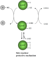
Catalytic mechanisms of Cys-GSTs. The deglutathionylation of GSH-conjugated substrates occurs via the nucleophilic attack of the catalytic cysteine which is assumed to be at least partially under the thiolate form at physiological pH, owing to a decreased pKa value. Consequently, the catalytic cysteine is itself glutathionylated and it is regenerated using a glutathione molecule. For Cys-GSTs having another cysteine either in the active site (some DHAR isoforms) or at proximity (some GSTL isoforms), the identification of proteins with an intramolecular disulfide suggests that this might constitute either an intermediate step of the catalytic mechanism or more likely a protective mechanism that prevents oxidation of the catalytic cysteine into sulfenic acid forms or eventually higher oxidized forms as sulfinic or sulfonic acid forms. In the case of the formation of a disulfide an additional glutathione molecule would be required. It may be that thioredoxin participate to this reduction step as DHAR was isolated as a thioredoxin targets.

References

    1. Aceto A., Sacchetta P., Dragani B., Bucciarelli T., Angelucci S., Longo V., et al. (1993). Glutathione transferase isoenzymes in olfactory and respiratory epithelium of cattle. Biochem. Pharmacol. 46, 2127–2133 10.1016/0006-2952(93)90600-2 - DOI - PubMed
    1. Allocati N., Casalone E., Masulli M., Ceccarelli I., Carletti E., Parker M. W., et al. (1999). Functional analysis of the evolutionarily conserved proline 53 residue in Proteus mirabilis glutathione transferase B1-1. FEBS Lett. 445, 347–350 10.1016/S0014-5793(99)00147-7 - DOI - PubMed
    1. Allocati N., Casalone E., Masulli M., Polekhina G., Rossjohn J., Parker M. W., et al. (2000). Evaluation of the role of two conserved active-site residues in beta class glutathione S-transferases. Biochem. J. 351 Pt 2, 341–346 10.1042/0264-6021:3510341 - DOI - PMC - PubMed
    1. Allocati N., Federici L., Masulli M., Di Ilio C. (2009). Glutathione transferases in bacteria. FEBS J. 276, 58–75 10.1111/j.1742-4658.2008.06743.x - DOI - PubMed
    1. Allocati N., Federici L., Masulli M., Favaloro B., Di Ilio C. (2008). Cysteine 10 is critical for the activity of Ochrobactrum anthropi glutathione transferase and its mutation to alanine causes the preferential binding of glutathione to the H-site. Proteins 71, 16–23 10.1002/prot.21835 - DOI - PubMed