Role of auxin during intercellular infection of Discaria trinervis by Frankia
- PMID: 25191330
- PMCID: PMC4139986
- DOI: 10.3389/fpls.2014.00399
Role of auxin during intercellular infection of Discaria trinervis by Frankia
Abstract
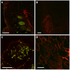
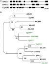
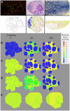
Nitrogen-fixing nodules induced by Frankia in the actinorhizal plant Discaria trinervis result from a primitive intercellular root invasion pathway that does not involve root hair deformation and infection threads. Here, we analyzed the role of auxin in this intercellular infection pathway at the molecular level and compared it with our previous work in the intracellular infected actinorhizal plant Casuarina glauca. Immunolocalisation experiments showed that auxin accumulated in Frankia-infected cells in both systems. We then characterized the expression of auxin transporters in D. trinervis nodules. No activation of the heterologous CgAUX1 promoter was detected in infected cells in D. trinervis. These results were confirmed with the endogenous D. trinervis gene, DtAUX1. However, DtAUX1 was expressed in the nodule meristem. Consistently, transgenic D. trinervis plants containing the auxin response marker DR5:VENUS showed expression of the reporter gene in the meristem. Immunolocalisation experiments using an antibody against the auxin efflux carrier PIN1, revealed the presence of this transporter in the plasma membrane of infected cells. Finally, we used in silico cellular models to analyse auxin fluxes in D. trinervis nodules. Our results point to the existence of divergent roles of auxin in intercellularly- and intracellularly-infected actinorhizal plants, an ancestral infection pathways leading to root nodule symbioses.
Keywords: AUX1; PIN; actinorrhiza; auxin; endosymbiosis; model; nitrogen fixation; systems biology.
Figures


Similar articles
-
Regulation of nodulation in the absence of N2 is different in actinorhizal plants with different infection pathways.J Exp Bot. 2003 Apr;54(385):1253-8. doi: 10.1093/jxb/erg131. J Exp Bot. 2003. PMID: 12654876
-
Cell remodeling and subtilase gene expression in the actinorhizal plant Discaria trinervis highlight host orchestration of intercellular Frankia colonization.New Phytol. 2018 Aug;219(3):1018-1030. doi: 10.1111/nph.15216. Epub 2018 May 23. New Phytol. 2018. PMID: 29790172
-
Diffusible factors from Frankia modify nodulation kinetics in Discaria trinervis, an intercellular root-infected actinorhizal symbiosis.Funct Plant Biol. 2011 Sep;38(9):662-670. doi: 10.1071/FP11015. Funct Plant Biol. 2011. PMID: 32480921
-
Actinorhizal symbioses and their N2 fixation.New Phytol. 1997 Jul;136(3):375-405. doi: 10.1046/j.1469-8137.1997.00755.x. New Phytol. 1997. PMID: 33863007 Review.
-
The diversity of actinorhizal symbiosis.Protoplasma. 2012 Oct;249(4):967-79. doi: 10.1007/s00709-012-0388-4. Epub 2012 Mar 8. Protoplasma. 2012. PMID: 22398987 Review.
Cited by
-
Accumulation of and Response to Auxins in Roots and Nodules of the Actinorhizal Plant Datisca glomerata Compared to the Model Legume Medicago truncatula.Front Plant Sci. 2019 Sep 24;10:1085. doi: 10.3389/fpls.2019.01085. eCollection 2019. Front Plant Sci. 2019. PMID: 31608077 Free PMC article.
-
The auxin phenylacetic acid induces NIN expression in the actinorhizal plant Datisca glomerata, whereas cytokinin acts antagonistically.PLoS One. 2025 Feb 3;20(2):e0315798. doi: 10.1371/journal.pone.0315798. eCollection 2025. PLoS One. 2025. PMID: 39899489 Free PMC article.
-
A Homeotic Mutation Changes Legume Nodule Ontogeny into Actinorhizal-Type Ontogeny.Plant Cell. 2020 Jun;32(6):1868-1885. doi: 10.1105/tpc.19.00739. Epub 2020 Apr 10. Plant Cell. 2020. PMID: 32276984 Free PMC article.
-
Recent advances in actinorhizal symbiosis signaling.Plant Mol Biol. 2016 Apr;90(6):613-22. doi: 10.1007/s11103-016-0450-2. Epub 2016 Feb 12. Plant Mol Biol. 2016. PMID: 26873697 Review.
-
Recent Advances in Adventitious Root Formation in Chestnut.Plants (Basel). 2020 Nov 11;9(11):1543. doi: 10.3390/plants9111543. Plants (Basel). 2020. PMID: 33187282 Free PMC article. Review.
References
-
- Berry A. M., Kahn R. K., Booth M. C. (1989). Identification of indole compounds secreted by Frankia HFPArI3 in defined culture medium. Plant Soil 118, 205–209 10.1007/BF02232808 - DOI
-
- Boisson-Dernier A., Chabaud M., Garcia F., Bécard G., Rosenberg C., Barker D. G. (2001). Agrobacterium rhizogenes-transformed roots of Medicago truncatula for the study of nitrogen-fixing and endomycorrhizal symbiotic associations. Mol. Plant. Microbe Interact. 14, 695–700 10.1094/MPMI.2001.14.6.695 - DOI - PubMed
LinkOut - more resources
Full Text Sources
Other Literature Sources
Miscellaneous

