

Chest CT Manifestations in Children with CVID: A 10-Year Report
- PMID: 25191439
- PMCID: PMC4153219
Chest CT Manifestations in Children with CVID: A 10-Year Report
Abstract
Background: This study aimed at evaluating HRCT pulmonary manifestations in children with Common Variable Immunodeficiency (CVID) hospitalized in the Pediatric Ward of Masih Daneshvari Hospital during a 10-year period.
Materials and methods: This retrospective study evaluated 25 children hospitalized with the diagnosis of CVID in the Pediatric Ward of Masih Daneshvari Hospital from 2001 to 2011 and their pulmonary HRCT scans were evaluated.
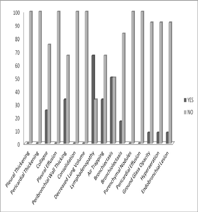
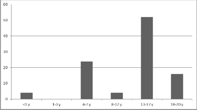
Results: The most common pulmonary HRCT findings were lymphadenopathy (66.7%), bronchiectasis (50%), air trapping (33.3%) and peribronchial wall thickening (33.3%). The highest percentage of CT-scan findings was detected in patients aged 13-17 yrs.
Conclusion: Most of the pulmonary changes due to CVID are preventable or treatable. Also, it is possible to prevent irreversible complications of disease if it is diagnosed early. Therefore, HRCT is strongly recommended as an accurate and effective method for monitoring and fast recognition of pulmonary manifestations of the disease especially bronchiectasis which is a very common finding indicative of poor prognosis.
Keywords: CT scan; Common variable immunodeficiency (CVID); HRCT.
Figures
References
-
- Bondioni MP, Duse M, Plebani A, Soresina A, Notarangelo LD, Berlucchi M, et al. Pulmonary and sinusal changes in 45 patients with primary immunodeficiencies: computed tomography evaluation. J Comput Assist Tomogr. 2007;31(4):620–8. - PubMed
-
- Cunningham-Rundles C, Bodian C. Common variable immunodeficiency: clinical and immunological features of 248 patients. Clin Immunol. 1999;92(1):34–48. - PubMed
-
- Bierry G, Boileau J, Barnig C, Gasser B, Korganow AS, Buy X, et al. Thoracic manifestations of primary humoral immunodeficiency: a comprehensive review. Radiographics. 2009;29(7):1909–20. - PubMed
-
- Cunningham-Rundles C. Common variable immunodeficiency. Curr Allergy Asthma Rep. 2001;1(5):421–9. - PubMed
-
- Oksenhendler E, Gérard L, Fieschi C, Malphettes M, Mouillot G, Jaussaud R, et al. Infections in 252 patients with common variable immunodeficiency. Clin Infect Dis. 2008;46(10):1547–54. - PubMed
LinkOut - more resources
Full Text Sources