. 2014 Aug 31:9:2.
doi: 10.1186/1747-1028-9-2. eCollection 2014.

Smurf2 E3 ubiquitin ligase modulates proliferation and invasiveness of breast cancer cells in a CNKSR2 dependent manner

Affiliations

Smurf2 E3 ubiquitin ligase modulates proliferation and invasiveness of breast cancer cells in a CNKSR2 dependent manner

Diana David et al. Cell Div. .

Abstract

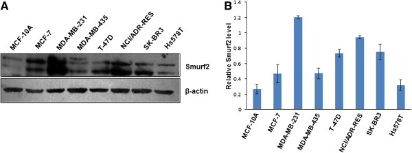
Background: Smurf2 is a member of the HECT family of E3 ubiquitin ligases that play important roles in determining the competence of cells to respond to TGF- β/BMP signaling pathway. However, besides TGF-β/BMP pathway, Smurf2 regulates a repertoire of other signaling pathways ranging from planar cell polarity during embryonic development to cell proliferation, migration, differentiation and senescence. Expression of Smurf2 is found to be dysregulated in many cancers including breast cancer. The purpose of the present study is to examine the effect of Smurf2 knockdown on the tumorigenic potential of human breast cancer cells emphasizing more on proliferative signaling pathway.

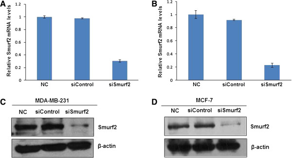
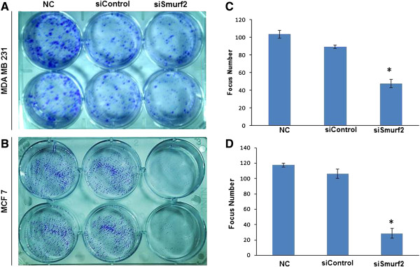
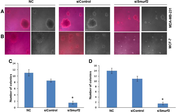
Methods: siRNAs targeting different regions of the Smurf2 mRNA were employed to knockdown the expression of Smurf2. The biological effects of synthetic siRNAs on human breast cancer cells were investigated by examining the cell proliferation, migration, invasion, focus formation, anchorage-independent growth, cell cycle arrest, and cell cycle and cell proliferation related protein expressions upon Smurf2 silencing.

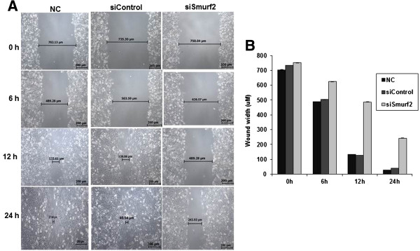
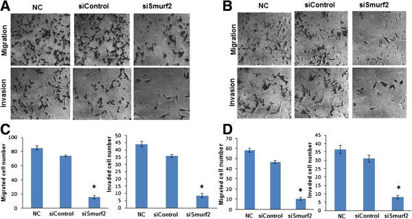
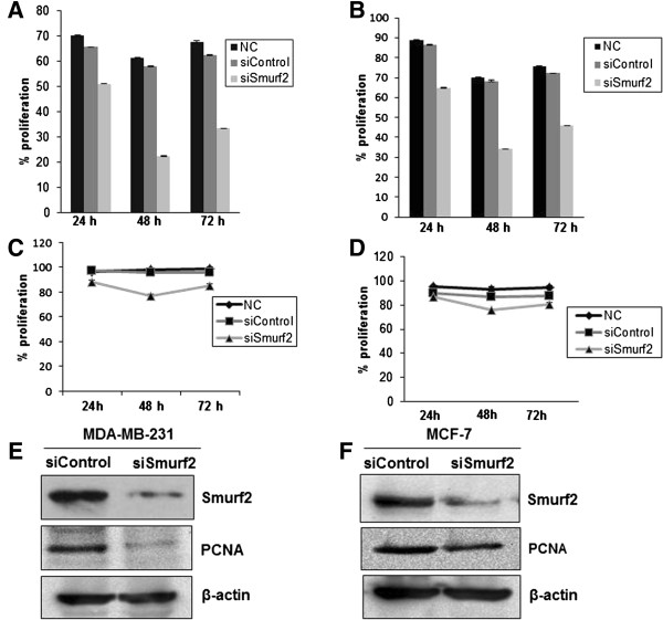
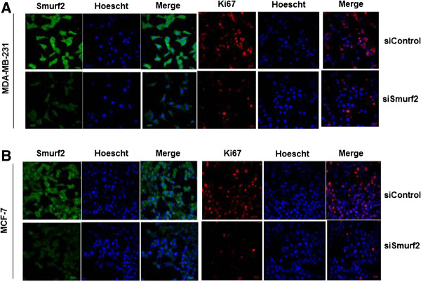
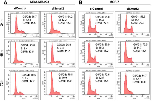
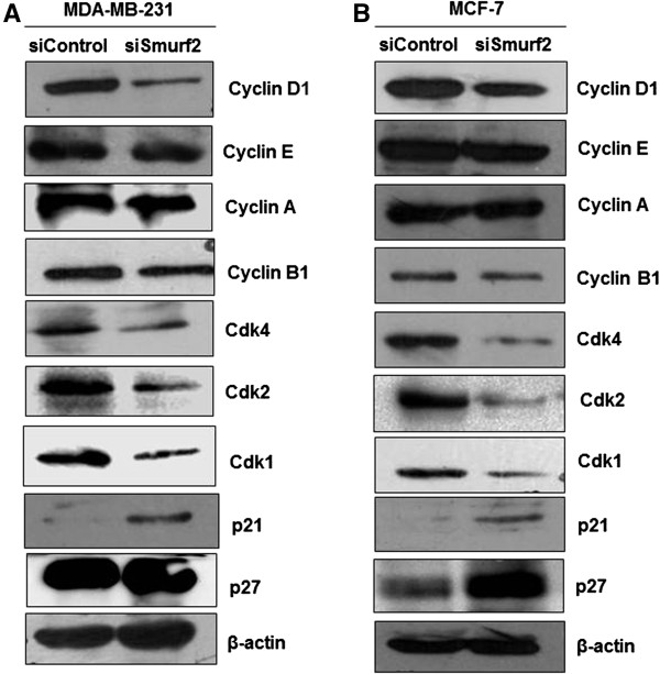
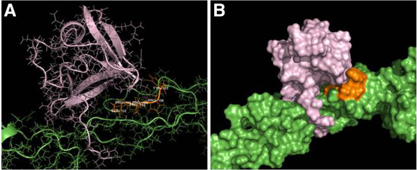
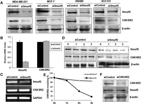
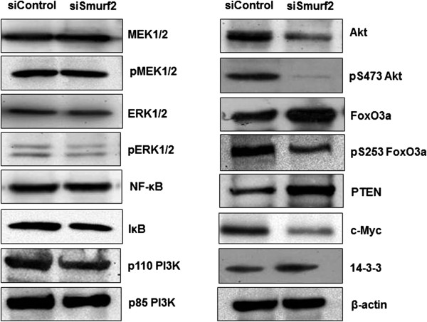
Results: Smurf2 silencing in human breast cancer cells resulted in a decreased focus formation potential and clonogenicity as well as in vitro cell migration/invasion capabilities. Moreover, knockdown of Smurf2 suppressed cell proliferation. Cell cycle analysis showed that the anti-proliferative effect of Smurf2 siRNA was mediated by arresting cells in the G0/G1 phase, which was caused by decreased expression of cyclin D1and cdk4, followed by upregulation p21 and p27. Furthermore, we demonstrated that silencing of Smurf2 downregulated the proliferation of breast cancer cells by modulating the PI3K- PTEN-AKT-FoxO3a pathway via the scaffold protein CNKSR2 which is involved in RAS-dependent signaling pathways. The present study provides the first evidence that silencing Smurf2 using synthetic siRNAs can regulate the tumorigenic properties of human breast cancer cells in a CNKSR2 dependent manner.

Conclusions: Our results therefore suggest a novel relation between Smurf2 and CNKSR2 thereby regulating AKT-dependent cell proliferation and invasion. Owing to the fact that PI3K-AKT signaling is hyperactivated in various human cancers and that Smurf2 also regulates cellular transformation, our results indicate that Smurf2 may serve as a potential molecule for targeted cancer therapy of certain tumour types including breast cancer.

Keywords: Breast cancer; CNKSR2; Oncogenic signaling; PI3K-AKT; Proliferation; Smurf2.


Figures

Figure 1
Figure 1
Smurf2 is upregulated in human breast cancer cell lines. (A) Smurf2 was found to be specifically upregulated in MDA-MB-231 cell line compared to other breast cancer cell lines. An untransformed immortalized cell line, MCF-10A was used as the control. β-actin was used to verify equal gel loading. (B) The bar graph indicates relative levels for Smurf2 protein in cancer cell lines to that in MCF10A. The density of each Smurf2 signal was normalized by β-actin. Data shows mean value ± S.E. from three independent experiments.
Figure 2
Figure 2
Knockdown effect of Smurf2 siRNA in MDA-MB-231 and MCF-7 cells. (A) MDA-MB-231 cells were transfected with Smurf2 siRNA (siSmurf2) and control siRNA (siControl) at a concentration of 80 pmols. Cells were harvested 36 hours after the transfection, and the silencing effect at the Smurf2 mRNA level was determined using real-time RT-PCR. (B) MCF-7 cells were transfected with Smurf2 siRNA, and control siRNA. The silencing effect at the Smurf2 mRNA level was measured using real-time RT-PCR. The silencing effect of Smurf2 siRNA at the protein level was determined 48 hours post-transfection using western blot in (C) MDA-MB-231 cells and (D) MCF-7 cells.
Figure 3
Figure 3
Silencing of Smurf2 inhibits focus formation. Forty-eight hours after the siRNA transfection, (A) MDA-MB-231 cells and (B) MCF-7 cells were seeded in six-well plates, and the medium was changed every 2 days. Cells cultured for 14 days were washed twice with 1xPBS, fixed by 4% paraformaldehyde, and stained with 0.5% crystal violet. Images of the colonies were obtained with a digital camera. (C), (D) Foci containing ≥50 cells were counted for MDA-MB-231 and MCF-7 cells and the result represented as mean ± standard deviation (n = 3). *P < 0.05 compared with control siRNA. NC-Negative control without siRNA.
Figure 4
Figure 4
Smurf2 silencing inhibits colony formation. A soft agar assay was conducted to examine the colony formation ability of (A) MDA-MB-231 cells and (B) MCF-7 cells. Twenty-four hours after the siRNA transfection, MDA-MB-231 and MCF-7 cells were seeded in 0.35% agarose in DMEM medium supplemented with 10% FBS at a density of 1 × 103 per 35-mm culture dish and allowed to grow for 21 days. The dishes were stained with 0.01% crystal violet, and the colonies were examined with microscope. Results are representative picture of colonies of two independent experiments done in triplicate. (C), (D) The number of colonies of MDA-MB-231 and MCF-7 cells were counted and the result represented as mean ± standard deviation (n = 3). *P < 0.05 compared with control siRNA. NC-Negative control without siRNA.
Figure 5
Figure 5
Smurf2 knockdown impairs cell motility. (A) Wound healing assay was done to evaluate the migration potential of MDA-MB-231 cells after silencing Smurf2 expression. Fifty-six hours after the transfection of siRNA, cells were wounded and monitored with a microscope every 6 hours. (B) The migration was determined by the rate of cells filling the scratched area and the result represented as mean ± standard deviation (n = 3). NC-Negative control without siRNA.
Figure 6
Figure 6
Silencing of Smurf2 expression inhibits migration and invasion abilities of MDA-MB-231 and MCF-7 cells. Cell migration was determined using Boyden transwell chambers. Forty-eight hours after the transfection with siRNA, (A) MDA-MB-231 cells and (B) MCF-7 cells were suspended in serum-free medium and seeded on 24-well transwell plates. Cells migrated though pores to the bottom surface of the transwell were fixed with with 10% formaldehyde, and stained with 0.5% crystal violet and counted. Six random microscopic fields were counted for each group. Cell invasion was assayed in transwell coated with Matrigel. Invaded cells were fixed, stained and counted. Six random microscopic fields were counted for each group. All experiments were performed in triplicates and repeated three times. Significant reduction of migration and invasion was observed after silencing Smurf2 expression in MDA-MB-231 and MCF-7 cells. (C), (D) The number of migrated or invaded cells of MDA-MB-231 and MCF-7 were counted from five or six randomly selected fields in a blind way and the result represented as mean ± standard deviation (n = 3). *P < 0.05 compared with control siRNA. NC, negative control without siRNA.
Figure 7
Figure 7
Smurf2 knockdown downregulates cell proliferation. Smurf2 knockdown decreases proliferation of MDA-MB-231 and MCF-7 cells which was measured with an MTT [3-(4,5-dimethylthiazol-2-yl)-2,5-diphenyl tetrazolium bromide] assay (A, B) and BrdU (5-bromo-2′-deoxyuridine) cell proliferation assay (C, D). Result represented as mean ± standard deviation (n = 3). The inhibition effect on cell proliferation is more significant at 48 hours rather than 72 hours posttransfection, after which proliferation gradually increased, probably due to transient transfection. Expression of PCNA (Proliferating cell nuclear antigen) was found to be downregulated in Smurf2 knockdown cells of (E) MDA-MB-231 and (F) MCF-7, 48 hours posttransfection.
Figure 8
Figure 8
Silencing of Smurf2 downregulates Ki-67 expression. Smurf2 knockdown significantly downregulated the expression of the proliferation marker Ki-67 in (A) MDA-MB-231 and (B) MCF-7 cells compared to control siRNA treated cells.
Figure 9
Figure 9
Silencing of Smurf2 leads to G0/G1 phase arrest. Cell cycle distribution of (A) MDA-MB-231 cells and (B) MCF-7 cells treated with Smurf2 siRNA and control siRNA were accessed by flow cytometry at 24, 48, and 72 hours post-transfection. Results are representative histogram of three independent experiments, plotting cell count vs. DNA content.
Figure 10
Figure 10
Expression levels of cell cycle regulatory proteins following Smurf2 knockdown. Silencing Smurf2 expression induces accumulation of cells in the G0/G1 phase. Lysates from (A) MDA-MB-231 and (B) MCF-7 cells following Smurf2 siRNA treatment were probed with the indicated antibodies. β-actin was used as the loading control.
Figure 11
Figure 11
Smurf2-WW2 domain interacts with CNKSR2. (A), (B) Docking of Smurf2-WW2/3 domains (purple coloured) and CNKSR2 (green coloured) using PATHDOCK and GROMACS indicate that the Smurf2-WW2 domain shows a better penetration and more stabilization with ‘SPPPPY’ motif (orange coloured) at 702–707 sequence region of CNKSR2, than Smurf2-WW3 domain.
Figure 12
Figure 12
Depletion of Smurf2 accelerates degradation of CNKSR2. (A) Depletion of Smurf2 by siRNA (siSmurf2) leads to decreased CNKSR2 protein levels in MDA-MB-231, MCF-7, SW480, and SCC131 cells. (B) Smurf2 does not affect CNKSR2 transcript level. RNA samples from MDA-MB-231 cells transfected with siSmurf2 were analyzed by qRT-PCR and further by (C) RT-PCR for the indicated transcripts. (D) MDA-MB-231 cells transfected with control siRNA and Smurf2 siRNA were treated with cycloheximide at 100 μg/ml, 48 hours post-transfection. Cells were harvested 0, 1, 2, or 3 h after cycloheximide addition and the cell lysate was subjected to SDS-PAGE followed by immunoblotting with specific antibodies. (E) Plot of CNKSR2 degradation rate was shown in the panel. (F) CNKSR2 depletion does not affect Smurf2 levels.
Figure 13
Figure 13
Smurf2 knockdown modulates the PI3K-AKT signaling pathway in a CNKSR2 dependent manner. Smurf2 knockdown diminishes Akt phosphorylation and FoxO3a-dependent cell proliferation. Lysates from control siRNA and Smurf2 siRNA treated MDA-MB-231 cells were probed with the indicated antibodies. β-actin was used as the loading control.

Similar articles

Cited by

References

    1. Osmundson EC, Ray D, Moore FE, Gao Q, Thomsen GH, Kiyokawa H. The HECT E3 ligase Smurf2 is required for Mad2-dependent spindle assembly checkpoint. J Cell Biol. 2008;183:267–277. doi: 10.1083/jcb.200801049. - DOI - PMC - PubMed
    1. Nakayama KI, Nakayama K. Ubiquitin ligases: cell-cycle control and cancer. Nat Rev Cancer. 2006;6:369–381. doi: 10.1038/nrc1881. - DOI - PubMed
    1. Kavsak P, Rasmussen RK, Causing CG, Bonni S, Zhu H, Thomsen GH, Wrana JL. Smad7 binds to Smurf2 to form an E3 ubiquitin ligase that targets the TGF beta receptor for degradation. Mol Cell. 2000;6:1365–1375. doi: 10.1016/S1097-2765(00)00134-9. - DOI - PubMed
    1. Moore FE, Osmundson EC, Koblinski J, Pugacheva E, Golemis EA, Ray D, Kiyokawa H. The WW-HECT protein Smurf2 interacts with the docking protein NEDD9/HEF1 for aurora a activation. Cell Div. 2010;5:22. doi: 10.1186/1747-1028-5-22. - DOI - PMC - PubMed
    1. Blank M, Tang Y, Yamashita M, Burkett SS, Cheng SY, Zhang YE. A tumor suppressor function of Smurf2 associated with controlling chromatin landscape andgenome stability through RNF20. Nat Med. 2012;18:227–234. doi: 10.1038/nm.2596. - DOI - PMC - PubMed