. 2014 Sep 5;9(9):e106569.
doi: 10.1371/journal.pone.0106569. eCollection 2014.

Evolution and development of ventricular septation in the amniote heart

Affiliations

Evolution and development of ventricular septation in the amniote heart

Robert E Poelmann et al. PLoS One. .

Abstract

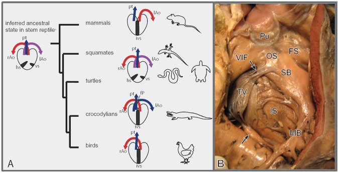
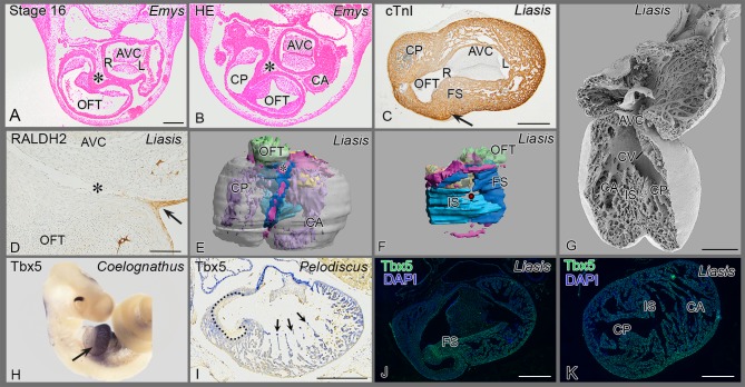
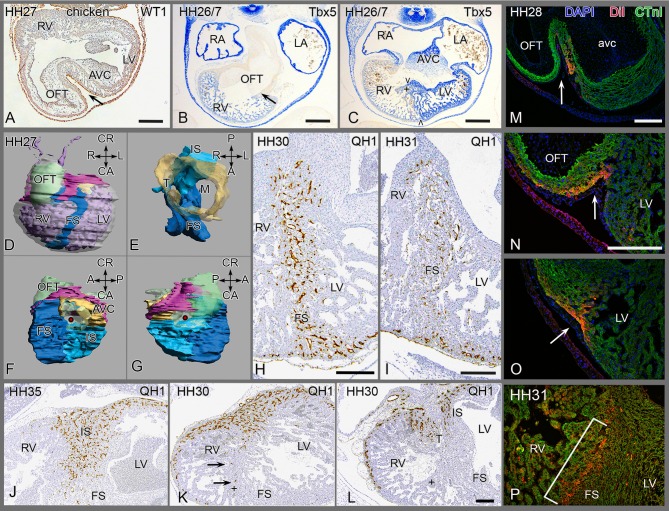
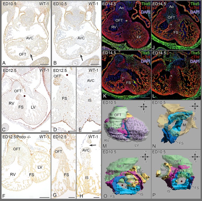
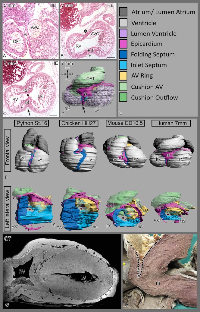
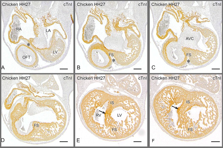
During cardiogenesis the epicardium, covering the surface of the myocardial tube, has been ascribed several functions essential for normal heart development of vertebrates from lampreys to mammals. We investigated a novel function of the epicardium in ventricular development in species with partial and complete septation. These species include reptiles, birds and mammals. Adult turtles, lizards and snakes have a complex ventricle with three cava, partially separated by the horizontal and vertical septa. The crocodilians, birds and mammals with origins some 100 million years apart, however, have a left and right ventricle that are completely separated, being a clear example of convergent evolution. In specific embryonic stages these species show similarities in development, prompting us to investigate the mechanisms underlying epicardial involvement. The primitive ventricle of early embryos becomes septated by folding and fusion of the anterior ventricular wall, trapping epicardium in its core. This folding septum develops as the horizontal septum in reptiles and the anterior part of the interventricular septum in the other taxa. The mechanism of folding is confirmed using DiI tattoos of the ventricular surface. Trapping of epicardium-derived cells is studied by transplanting embryonic quail pro-epicardial organ into chicken hosts. The effect of decreased epicardium involvement is studied in knock-out mice, and pro-epicardium ablated chicken, resulting in diminished and even absent septum formation. Proper folding followed by diminished ventricular fusion may explain the deep interventricular cleft observed in elephants. The vertical septum, although indistinct in most reptiles except in crocodilians and pythonidsis apparently homologous to the inlet septum. Eventually the various septal components merge to form the completely septated heart. In our attempt to discover homologies between the various septum components we aim to elucidate the evolution and development of this part of the vertebrate heart as well as understand the etiology of septal defects in human congenital heart malformations.


Conflict of interest statement

Competing Interests: The authors have declared that no competing interests exist.

Figures

Figure 1
Figure 1. Evolution and septation of the heart.
A. Evolution of hearts in higher vertebrates. Archosaurs (crocodilians, birds) and mammals independently evolved complete ventricular septation. Birds and mammals have lost either a left (lAo) or right (rAo) aorta. The horizontal (hs) and vertical septum (vs) are schematically indicated, together with the pulmonary trunk (Pt). The evolutionary tree is based on ref (2). B. Septum components in the human heart. Right face of the septum in a human heart after opening the right ventricle (RV), with inlet and folding components. Dissection line of the RV free wall in pink. Abbreviations: FS folding septum, IS inlet septum; MB moderator band; Pu pulmonary semilunar valve leaflets; SB septal band; TV anterior tricuspid valve leaflet with chordae tendineae (arrows) connected to SB and IS.; VIF ventriculo-infundibular fold. Fig. courtesy dr. L. Houyel.
Figure 2
Figure 2. Reptile cardiac development.
(A, B) The epicardial cushion (*) is located between OFT and AV cushions. (C, D) cardiac troponin I (cardiac muscle) and RALDH2 (epicardial cells) stainings show folding septum (arrow, asterisk). (E) 3D reconstruction in an anterior view, the epicardial patches are depicted in pink. See also Figure S1 1 for full animation. (F) right sided view of the septum, folding (FS) and inlet (IS) septum are depicted in shades of blue. For further colors see legend to Fig. 5E. (G) Scanning electron microscopy of anterior inner face, note communication between the three cava. The folding (syn. horizontal) septum is out of view. (H) A sharp decline of Tbx5 mRNA expression (arrow) between cavum dorsale and OFT. (I) Sharp boundary at muscular OFT (inside of dotted line) and wall of cavum pulmonale (outside dotted line), but the tip of trabeculations in the cavum dorsale stain strongly (arrows). (J) Section downstream of Fig C, showing sharp decline of Tbx5 protein expression at folding septum. (K) Section more to the apex of J, showing uniform immunostaining for Tbx5. Abbreviations as in Fig 1, others: AVC: AV cushions; ca, cp, cv: cavum arteriosum, pulmonale and venosum; L left AV orifice; OFT outflow tract cushions; R right AV orifice; →: infolding; * epicardium and EPDCs; • position of cavum venosum in 3D reconstruction of Fig F.
Figure 3
Figure 3. Development of chicken septum.
(A) epicardium (→) infolding, located between OFT and AVcanal. (B) In situ hybridisation showing weakly positive Tbx5 of the RV and negative OFT with boundary (arrow). The atria are strongly positive. (C) more posterior section of the same embryo through folding septum (FS), the stronger left sided expression is evident, as is the septal band (+); boundary (> <) indicates FS. (D-G) 3D reconstruction with septum components and epicardial cushion. See also figure S2 for full animation and Fig. 6 for underlying sections, explaining the various components. (H-L) PEO quail-chicken chimeras. (H, I) anterior quail PEO(+liver) transplant, quail endothelial cells are exclusively present in FS and anterior free wall (J-L) posterior PEO (+liver) transplant with quail vascular profiles in IS (J, K) and right face of tricuspid orifice (L), but not in FS. (K) Several quail cells (arrows) in septal band (+), but FS does not harbor quail cells and remains negative (K, L). (M-P) DiI marking at HH17 of anterior myocardium surviving until HH28 and 31. (M) parts of the DiI patch (arrow) after survival to HH28 on left, (N) DiI on the right face and (O) DiI near the apex. (P) DiI inside the septum at HH31. Abbrev. as in Fig 2. Others: AVC atrioventricular cushions; LA/LV left atrium and ventricle; RA/RV right atrium and ventricle; + septal band.
Figure 4
Figure 4. Septum formation in the mouse.
(A, B) Almost transverse sections of the same embryo showing WT1+ epicardial cells in the folding septum (FS,→) at ED 10.5, Fig. B is more apically located. (C, D) epicardial cells in FS of wildtype mouse at ED 12.5 and (E) present in the inlet septum underneath the posterior AV cushion. Note: WT1 staining of mesenchyme in septal OFT cushion is unrelated to epicardial cells. (F-H) Podoplanin mutant with diminutive PEO, presents with sparse epicardium lining the pericardial cavity (F, G) and with an underdeveloped septum lacking EPDCs in both FS (G) and inlet septum (IS) (H). (I-L) Immunostained for Tbx5 in a wild type mouse ED 14.5, four levels from anterior-posterior. (I) Tbx5 in LV trabeculations but not in the RV close to the outflow tract; core of septum is negative. (J-L) More posteriorly located sections, trabeculations in RV belonging to the inlet part become positive for Tbx5. (M-P) Four positions of a 3D Amira reconstruction of ED 10.5. The epicardial cushion in pink (*), the folding septum in dark blue and the inlet septum in light blue. Endocardial cushions in green and the AVC myocardium in yellow. See also Fig. S3 for animated 3D. Abbrev. AVC atrioventricular cushions; FS folding septum; IS inlet septum; LV left ventricle; M mitral orifice; RV right ventricle; OFT outflow tract cushion; T tricuspid orifice, • interventricular communication, + septal band.
Figure 5
Figure 5. Embryonic human and adult elephant hearts.
(Fig. A, B) Epicardial cushion (*) in inner curvature between OFT and AVC. The inlet septum becomes apparent more apically (Fig. C). (D) and Fig S4 represent a reconstructed 7 mm embryo with folding and inlet septum formation. (F) 3D Comparison of development. Top row, 4 species showing relation of epicardial cushion (pink) with folding septum (dark blue). Bottom row, left lateral view, showing connection of folding septum with the inlet septum (light blue). The interventricular foramen connects left and right ventricle. In python this connection is represented by the cavum venosum. The AV myocardium is depicted in yellow and the various cushion tissues in green. (G, H) adult elephant heart (G) CT-image of first bifid elephant heart. (H) Anatomical dissection of the interventricular septum of the second heart. Left descending coronary artery (arrow) and the deep epicardial fat pad are outlined. Note the relative absence of a folding component. The dissected chordae tendineae of the tricuspid valve are visible in the lower part of Fig 5H. Abbrev. AVC atrioventricular cushions; FS folding septum; IS inlet septum; LV left ventricle; RV right ventricle; OFT outflow tract cushion.
Figure 6
Figure 6. Chicken embryo HH27.
Provides 6 sections of a serially sectioned chicken embryo (HH27) to demonstrate the merging of the folding and inlet components before septation is finished. From this embryo Fig 3D and Fig. S2 have been reconstructed. Similar series served as basis for the other species depicted in Fig S1, S3 and S4. Fig 6 .A is most cranial, showing the epicardial cushion (*) at a level between outflow tract (OFT) and the right (RA) and left atria (LA) with the AV cushions in between. The cranial cap of the left ventricle (LV) is grazed in the section. Fig 6 .B and C give the cranial extension of the folding septum (FS) with the epicardial cushion (*) located between the outflow tract and the fused AV cushions (AVC). Fig 6 .D shows the FS bordering the interventricular foramen. It is evident that the core of the folding septum is lined on the left and right side by many trabeculations. Fig. 6.E The AV cushions are attached to the flanks of the inlet septum where also the tip of the septal OFT cushion is found (arrow). The inlet (IS) and folding components have fused and constitute the floor of the interventricular foramen. Fig 6 .E, F The right AV junction is present in the RV immediately above the arrow and can be traced upstream in Fig F and downstream in Fig. D. Note the close relationship to the IS. The folding septum becomes less compact and the trabeculations become more conspicuous. Abbrev. AVC atrioventricular cushions; FS folding septum; IS inlet septum; LA left atrium; LV left ventricle; RA right atrium; RV right ventricle; OFT outflow tract cushion with its proximal tip indicated by arrow in Fig. F.

Similar articles

Cited by

References

    1. Holmes EB (1975) A reconsideration of the phylogeny of the tetrapod heart. J Morphol 147: 209–228. - PubMed
    1. Wang Z, Pascual-Anaya J, Zadissa A, Li W, Niimura Y, et al. (2013) The draft genomes of soft-shell turtle and green sea turtle yield insights into the development and evolution of the turtle-specific body plan. Nat Genet 45: 701–706 10.1038/ng.2615 - DOI - PMC - PubMed
    1. Jacobs JP, Quintessenza JA, Burke RP, Mavroudis C (2000) Congenital Heart Surgery Nomenclature and Database Project: atrial septal defect. Ann Thorac Surg 69: S18–S24. - PubMed
    1. Gittenberger-de Groot AC, Vrancken Peeters M-PFM, Mentink MMT, Gourdie RG, Poelmann RE (1998) Epicardium-derived cells contribute a novel population to the myocardial wall and the atrioventricular cushions. Circ Res 82: 1043–1052. - PubMed
    1. Von Gise A, Pu WT (2012) Endocardial and epicardial epithelial to mesenchymal transitions in heart development and disease. Circ Res 110: 1628–1645 10.1161/CIRCRESAHA.111.259960 - DOI - PMC - PubMed

Publication types

Substances

LinkOut - more resources