. 2014 Sep 5;9(9):e106465.
doi: 10.1371/journal.pone.0106465. eCollection 2014.

A novel fatty acid-binding protein-like carotenoid-binding protein from the gonad of the New Zealand sea urchin Evechinus chloroticus

Affiliations

A novel fatty acid-binding protein-like carotenoid-binding protein from the gonad of the New Zealand sea urchin Evechinus chloroticus

Jodi Pilbrow et al. PLoS One. .

Abstract

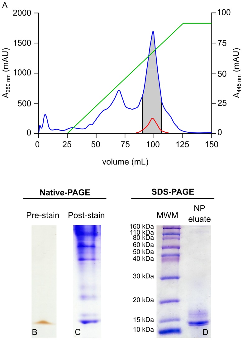
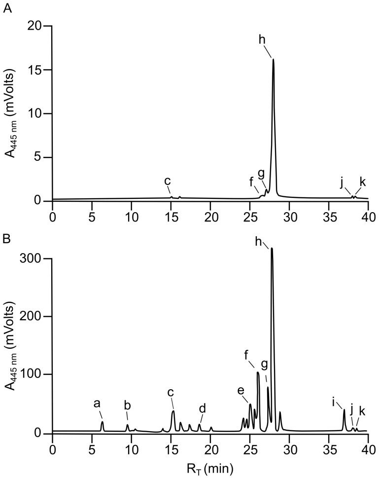
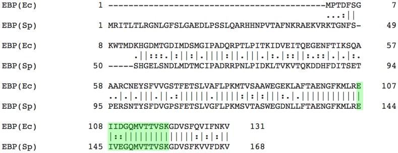
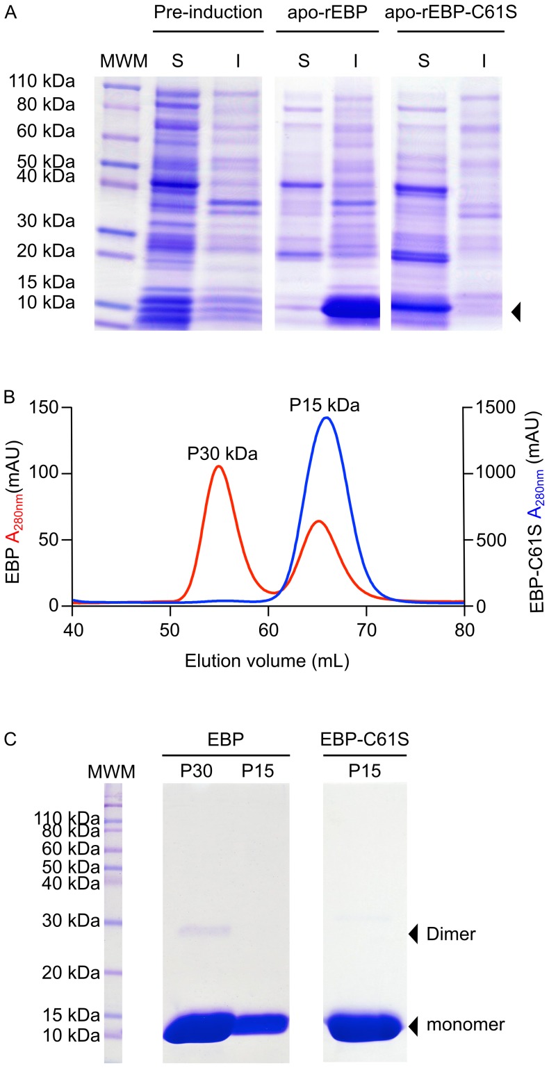
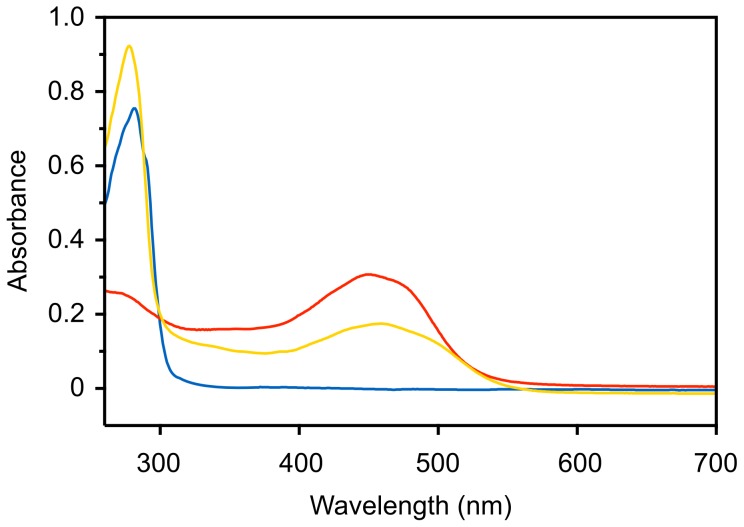
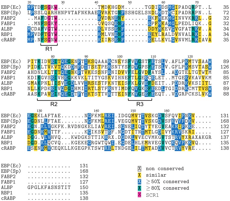
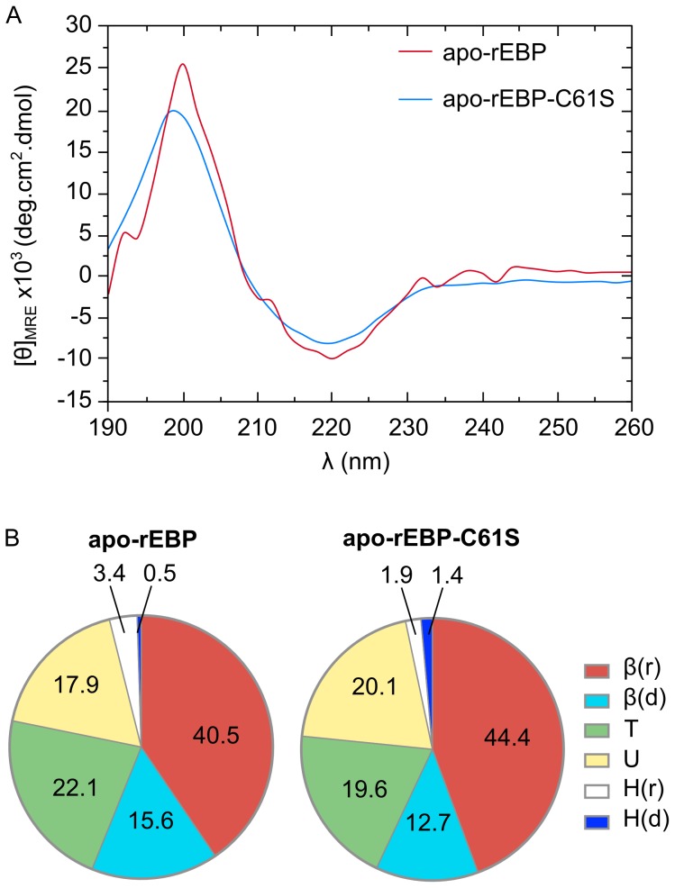
A previously uncharacterized protein with a carotenoid-binding function has been isolated and characterized from the gonad of the New Zealand sea urchin Evechinus chloroticus. The main carotenoid bound to the protein was determined by reversed phase-high performance liquid chromatography to be 9'-cis-echinenone and hence this 15 kDa protein has been called an echinenone-binding protein (EBP). Purification of the EBP in quantity from the natural source proved to be challenging. However, analysis of EBP by mass spectrometry combined with information from the Strongylocentrotus purpuratus genome sequence and the recently published E. chloroticus transcriptome database, enabled recombinant expression of wild type EBP and also of a cysteine61 to serine mutant that had improved solubility characteristics. Circular dichroism data and ab initio structure prediction suggests that the EBP adopts a 10-stranded β-barrel fold consistent with that of fatty acid-binding proteins. Therefore, EBP may represent the first report of a fatty acid-binding protein in complex with a carotenoid.


Conflict of interest statement

Competing Interests: The authors have declared that no competing interests exist.

Figures

Figure 1
Figure 1. Isolation of a putative CBP by 1D native-PAGE.
E. chloroticus gonad-soluble protein extract was fractionated by anion exchange chromatography, A. Gonad protein extract corresponding to 1 g wet weight gonad was loaded onto a 5 mL HiTrap Q-Sepharose column and bound protein was eluted by a 0–100% gradient of 1M NaCl (green line). The absorbance of the column effluent was monitored at 280 nm (blue) and 445 (red). Fractions absorbing at both 280 nm and 445 nm (grey zone) were pooled concentrated. B. A 20 µL aliquot of the concentrate was analyzed on 1D native-PAGE, shown prior to staining. C. A duplicate loading on 1D native-PAGE was stained with Coomassie blue R-250. D. The yellow/orange band visible on the pre-stained gel in B. was excised and the protein was eluted and then analyzed by 1D SDS-PAGE and stained with Coomassie blue R-250.
Figure 2
Figure 2. RP-HPLC of carotenoids extracted from EBP and from E. chloroticus gonad tissue.
Carotenoids extracted from native-PAGE eluate (Figure 1) A and from lyophilized whole gonad tissue B, were separated by RP-HPLC. Each carotenoid extraction corresponded to a whole gonad, of the same weight, taken from a single animal. For RP-HPLC analysis a 100 µL aliquot of carotenoid was injected onto a C30 RP-HPLC and carotenoids were eluted with a methanol/TBME gradient. Column effluent was monitored at 445 nm and carotenoids were identified by comparison of retention times (RT) to commercial standards. The carotenoids identified were a. fucoxanthinol, b. fucoxanthin, c. astaxanthin, d. canthaxanthin, e. lutein, f. isozeaxanthin, g. all-trans-echinenone, h. 9′-cis-echinenone, i. α-carotene, j. all-trans-β-carotene and k. 9′-cis-β-carotene.
Figure 3
Figure 3. Global pairwise alignment of EBP(Ec) and EBP(Sp) amino acid sequences.
A global pairwise alignment of EBP(Sp) and EBP(Ec) amino acid sequences was performed using EMBOSS Needle. The peptide sequence derived by mass spectrometry of an in-gel digest of EBP(Ec) is highlighted in green.
Figure 4
Figure 4. Expression and purification of recombinant apo-EBP and apo-EBP-C61S.
Apo-rEBP and apo-rEBP-C61S were produced using E.coli expression strain BL21(DE3), Expression levels were evaluated on reducing 1D SDS-PAGE, prior to induction and 6 h post-induction, A. Soluble (S) and insoluble (I) fractions of bacterial cell lysate are indicated. Arrowhead indicates the position of EBP and EBP-C61S bands, the identities of which were confirmed by mass spectrometry (Figure S1). Apo-rEBP and apo-rEBP-C61S were purified from the soluble fractions of the bacterial cell lysates. Gel-permeation chromatography was performed under non-reducing conditions for the final stage of purification on a 16/60 Sephacryl S-100 column, with the effluent monitored at 280 nm, B. Apo-rEBP is shown on the left y-axis (red) and apo-rEBP-C61S on the right y-axis (blue). The molecular weight of the eluting species was estimated with reference to the manufacturers Sephacryl S-100 column standard curve. The fractions within each peak were pooled and a 10 µL aliquot of each was analyzed on reducing 1D SDS-PAGE, C. The positions of the monomeric and dimeric forms of protein are indicated by black arrowheads.
Figure 5
Figure 5. Absorbance spectra of apo and holo-rEBP-C61S.
Following protein-ligand reconstitution and removal of unbound carotenoid the absorbance spectrum of holo-rEBP-C61S (1 mg.mL-1) was measured between 260 nm and 700 nm, against a reference solution of 20 mM HEPES pH 8.0 (yellow line). Spectra of 1 mg.mL-1 apo-rEBP-C61S in 20 mM HEPES pH 8.0 (blue line) and purified 9′-cis-echinenone at 0.9 mg.mL-1 in methanol (red line) are shown for comparison.
Figure 6
Figure 6. Multiple sequence alignment of EBP(Ec) and EBP(Sp) with members of the FABP family.
A multiple sequence alignment of EBP(Ec) and EBP(Sp) with five members of the FABP family was performed using MUSCLE. The five FABP sequences used in the alignment were; intestinal-FABP-2 (FABP2), liver-FABP-1 (FABP1), adipocyte lipid-binding protein (ALBP) from Rattus norvegicus, retinol-binding protein-1 (RBP1) and cellular retinoic acid-binding protein (cRABP) from Homo sapiens. Positions where ≥50% of residues are similar are shaded yellow. Positions where residue identity is ≥80% are shaded blue. Positions with ≥80% residue identity are shaded green. SCR1 (≥80% identity) is shaded magenta and the three regions of clustered ≥80% amino acid identity are labelled R1, R2 and R3. Amino acid position numbering is with respect to EBP(Sp), for which the first 20 amino acids are not shown.
Figure 7
Figure 7. Ab initio secondary structure prediction of EBP.
Comparison of secondary structure predictions for EBP from five different web-based servers; YASPIN, SPINEX, NetSurfP, PSIPRED and PHD. Blue shaded regions indicate β-strand structures, red shaded regions helical structures and unshaded regions, coils. The consensus prediction, defined as the prediction made by 3/5 or more of the servers, is displayed above the sequence. Blue arrows represent β-strand structures SA-J, red coils represent helical structures labeled H1-2 and gaps indicate coil regions. Black boxes indicate the amino acids that were ≥80% conserved between FABP sequences included in the MSA (figure 6).
Figure 8
Figure 8. CD spectra of recombinant apo-rEBP and apo-rEBP-C61S.
Spectra were collected in the presence of 5 mM phosphate, pH 7.5 at 18°C. A. CD spectra of apo-rEBP is shown in red and apo-rEBP-C61S is shown in blue. Predictions of secondary structural elements in apo-rEBP and apo-rEBP-C61S. B. The Δε data derived from CD spectra were analyzed using CDPro and percentage averages of each element, predicted by CDSSTR and CONTINLL, are shown for the SMP53 protein reference set. Regular helix - H(r), distorted helix - H(d), regular sheet - β(r), distorted sheet – β(d), turns - T and unordered – U.

References

    1. Guidice G (1973) Developmental biology of the sea urchin embryo. New YorkAcademic Press.
    1. McClay DR (2011) Evolutionary crossroads in developmental biology: sea urchins. Development 138: 2639–2648. - PMC - PubMed
    1. Ernst SG (2011) Offerings from an Urchin. Developmental Biology 358: 285–294. - PubMed
    1. Williams H (2002) Sea urchin fisheries of the world: A review of their status, management strategies and biology of the principal species. Department of Primary Industries, Water and Environment. Tasmania.
    1. Andrew NL, Agatsuma Y, Ballesteros E, Bazhin AG, Creaser EP, et al. (2002) Status and management of world sea urchin fisheries. Oceanography and Marine Biology Annual Review 20: 343–425.

Publication types

MeSH terms