. 2014 Nov;80(22):6976-91.
doi: 10.1128/AEM.01576-14. Epub 2014 Sep 5.

The fatty acid signaling molecule cis-2-decenoic acid increases metabolic activity and reverts persister cells to an antimicrobial-susceptible state

Affiliations

The fatty acid signaling molecule cis-2-decenoic acid increases metabolic activity and reverts persister cells to an antimicrobial-susceptible state

Cláudia N H Marques et al. Appl Environ Microbiol. 2014 Nov.

Abstract

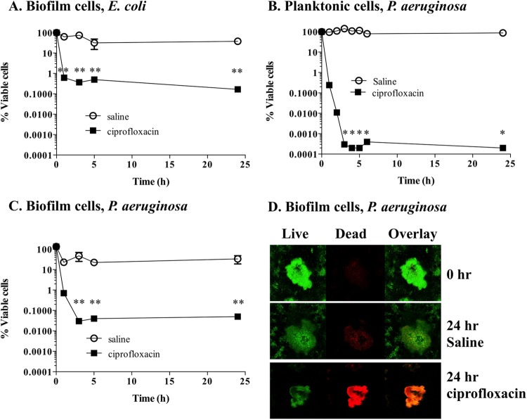
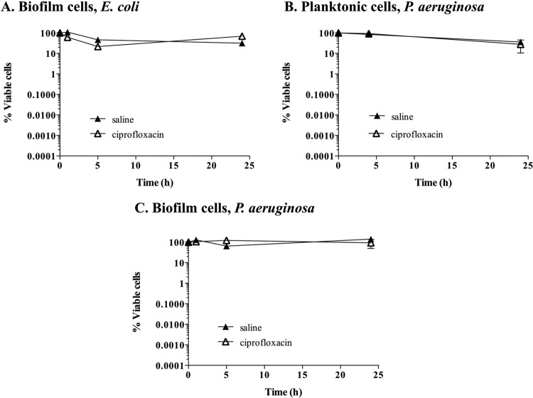
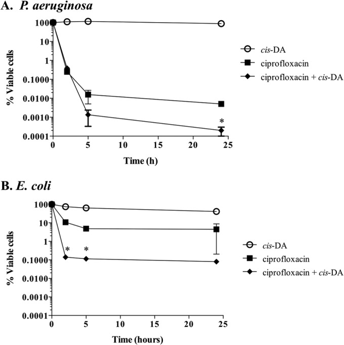
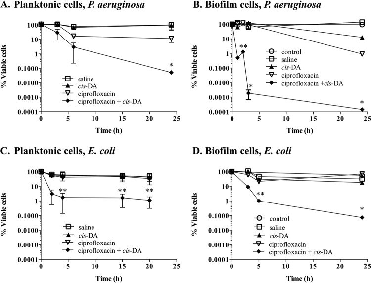
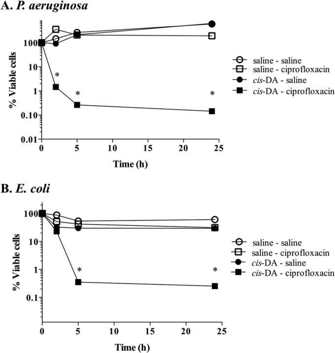
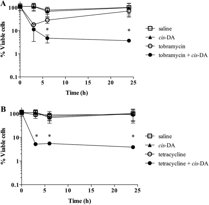
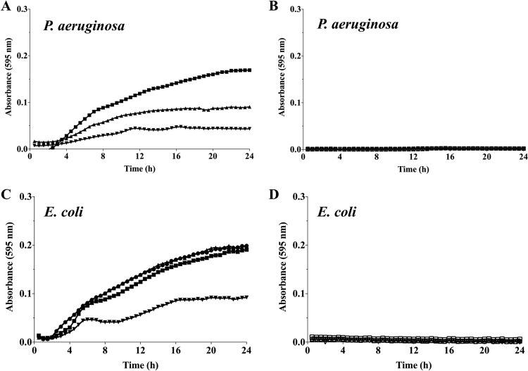
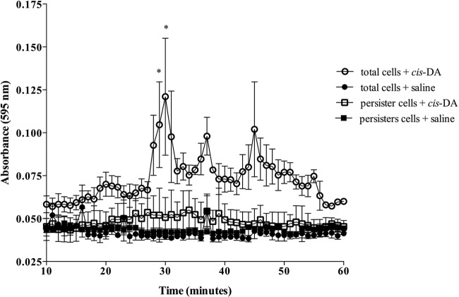
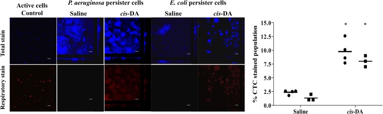
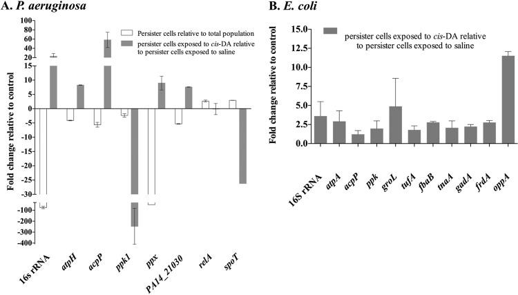
Persister cells, which are tolerant to antimicrobials, contribute to biofilm recalcitrance to therapeutic agents. In turn, the ability to kill persister cells is believed to significantly improve efforts in eradicating biofilm-related, chronic infections. While much research has focused on elucidating the mechanism(s) by which persister cells form, little is known about the mechanism or factors that enable persister cells to revert to an active and susceptible state. Here, we demonstrate that cis-2-decenoic acid (cis-DA), a fatty acid signaling molecule, is able to change the status of Pseudomonas aeruginosa and Escherichia coli persister cells from a dormant to a metabolically active state without an increase in cell number. This cell awakening is supported by an increase of the persister cells' respiratory activity together with changes in protein abundance and increases of the transcript expression levels of several metabolic markers, including acpP, 16S rRNA, atpH, and ppx. Given that most antimicrobials target actively growing cells, we also explored the effect of cis-DA on enhancing antibiotic efficacy in killing persister cells due to their inability to keep a persister cell state. Compared to antimicrobial treatment alone, combinational treatments of persister cell subpopulations with antimicrobials and cis-DA resulted in a significantly greater decrease in cell viability. In addition, the presence of cis-DA led to a decrease in the number of persister cells isolated. We thus demonstrate the ability of a fatty acid signaling molecule to revert bacterial cells from a tolerant phenotype to a metabolically active, antimicrobial-sensitive state.


Figures

FIG 1
FIG 1
Isolation of persister cells from planktonic and biofilm populations. Stationary-phase planktonic cultures and 6-day biofilms were exposed to saline or ciprofloxacin in saline for a period of 24 h. Cell viability was determined throughout the experiment. (A to C) E. coli biofilm populations (A), P. aeruginosa planktonic populations (B), and P. aeruginosa biofilm populations (C). (D) Cell viability of P. aeruginosa biofilms was also assessed by using SYTO9 (live cells) and propidium iodide (dead cells) at 0 h and at 24 h for saline- and ciprofloxacin-exposed cells. The averages of data from 3 experiments with 2 replicates per experiment are shown. Error bars indicate standard deviations (*, P < 0.001; **, P < 0.01 [significantly different from persister cells treated with saline, as indicated by one-way ANOVA]).
FIG 2
FIG 2
Confirmation of the persister cell state. Persister cells isolated from planktonic and biofilm populations were exposed to saline or ciprofloxacin in saline for 24 h. Cell viability was determined throughout the experiment. Shown are E. coli biofilm populations (A), P. aeruginosa planktonic populations (B), and P. aeruginosa biofilm populations (C). The averages of data from 3 experiments with 2 replicates per experiment are shown. Error bars indicate standard deviations.
FIG 3
FIG 3
Effect of cis-DA on the percent persister cell subpopulation. P. aeruginosa (A) and E. coli (B) persister cells were isolated from stationary-phase planktonic cultures in the presence and absence of cis-DA. Cultures were exposed to ciprofloxacin in saline (20 μg/ml), ciprofloxacin with cis-DA in saline, and cis-DA in saline for a period of 24 h. Cell viability was determined throughout the experiment. The averages of data from 3 experiments with 2 replicates per experiment are shown. Error bars indicate standard deviations (*, P < 0.01 [significantly different from persister cells treated with ciprofloxacin, as indicated by one-way ANOVA]).
FIG 4
FIG 4
Tolerance of persister cells to ciprofloxacin is reduced in the presence of cis-DA. Persister cell subpopulations of P. aeruginosa planktonic populations (A), P. aeruginosa biofilms (B), E. coli planktonic populations (C), and E. coli biofilms (D) were exposed to saline, cis-DA in saline, ciprofloxacin in saline, and ciprofloxacin with cis-DA in saline. Biofilm and planktonic populations were exposed to 150 μg/ml and 20 μg/ml of ciprofloxacin, respectively. Experiments were performed for a period of 24 h, and cell viability was determined throughout the experiment. Experiments with persister cells derived from biofilms had one additional control, a total biofilm population treated with cis-DA in saline (control). P. aeruginosa cells were exposed to 100 nM cis-DA, and E. coli cells were exposed to 310 nM cis-DA. The averages of data from 3 experiments are shown. Error bars indicate standard deviations (*, P < 0.001; **, P < 0.01 [significantly different from persister cells treated with ciprofloxacin, saline, and cis-DA, as indicated by one-way ANOVA]).
FIG 5
FIG 5
Effect of cis-DA on maintenance of the persister state. P. aeruginosa (A) and E. coli (B) persister cells were exposed to cis-DA in saline or saline for a period of 20 h prior to exposure to saline or ciprofloxacin in saline for a further 24 h. Viability was determined throughout the experiment. The averages of data from 3 experiments are shown. Error bars indicate standard deviations (*, P < 0.001 [significantly different from persister cells treated with saline, as indicated by one-way ANOVA]).
FIG 6
FIG 6
Tolerance of persister cells to tobramycin and tetracycline in the presence of cis-DA. Persister cell subpopulations of P. aeruginosa planktonic populations were exposed to tobramycin (A) and tetracycline (B) in the presence and absence of cis-DA (100 nM) for a period of 24 h. Shown are data for treatments with saline, cis-DA in saline, antimicrobial in saline, and antimicrobial and cis-DA in saline. Tetracycline was used at a concentration of 100 μg/ml, and tobramycin was used at a concentration of 20 μg/ml. Viability was determined throughout the experiment. The averages of data from 3 experiments are shown. Error bars indicate standard deviations (*, P < 0.01 [significantly different from persister cells treated with saline, cis-DA, and antimicrobials, as indicated by one-way ANOVA]).
FIG 7
FIG 7
Use of cis-DA as a carbon source. Planktonic cultures of P. aeruginosa and E. coli were grown in EPRI medium containing either glucose or cis-DA as a carbon source. The concentrations of glucose (closed symbols) (A and C) and cis-DA (open symbols) (B and D) used were 100 nM (▼/▽), 300 nM (▲/△), and 1,000 nM (◼/◻). Results correspond to representative data from 3 experiments with a total of at least 10 biological replicates.
FIG 8
FIG 8
Persister cells do not disperse in the presence of cis-DA. Biofilms were composed of total cell populations (exposed to saline for a period of 18 h) or persister cell populations (exposed to ciprofloxacin at 150 μg/ml for 18 h). Biofilms were exposed to cis-DA (100 nM) in saline or saline for a period of 60 min, and dispersion was evaluated by measuring the absorbance at a wavelength of 595 nm. The averages of data from 3 experiments are shown. Error bars indicate standard deviations (*, P < 0.01 [significantly different from persister cells treated with saline, as indicated by one-way ANOVA]).
FIG 9
FIG 9
Respiratory activity of persister cells upon exposure to cis-DA. Respiratory activity of P. aeruginosa and E. coli biofilm persister cells was evaluated by the ability of the cells to metabolize CTC into fluorescent formazan following 1 h of incubation with saline or cis-DA in saline. SYTO40 was used to stain the overall population. The percentage of the CTC-stained population of persister cells compared to the SYTO40-stained population was calculated. Error bars indicate standard deviations. Symbols in the graph correspond to P. aeruginosa (●) and E. coli (■). Bar = 25 μm (*, P < 0.001 [significantly different from persister cells treated with saline alone, as indicated by one-way ANOVA]).
FIG 10
FIG 10
Transcription abundance of metabolic activity markers. Persister cells were selected from biofilm cultures of P. aeruginosa (A) and E. coli (B). The relative expression levels of the metabolic activity markers 16S rRNA, acpP, atpH, atpA, ppk, and ppx were evaluated upon exposure of persister cells to cis-DA in saline and compared to the levels upon exposure to saline alone. P. aeruginosa was exposed to cis-DA for a period of 1 h, while E. coli was exposed for a period of 6 h. The relative expression level for P. aeruginosa persister cells derived from biofilms was also compared to that for the overall nonpersister population. In addition, the relative expression levels of relA and spoT, known to be involved in persister cell formation, were also evaluated for P. aeruginosa. Genes related to some proteins present in higher abundance upon exposure of persister cells were also evaluated. The CT value of the housekeeping gene mreB remained constant throughout the different treatments (P > 0.5 by ANOVA and no difference between treatments by Tukey's multiple-comparison test). Experiments were carried out at least in triplicate. Error bars denote standard deviations. A significant change was considered to occur when a 2-fold change in the relative expression level occurred.

Similar articles

Cited by

References

    1. Balaban NQ, Merrin J, Chait R, Kowalik L, Leibler S. 2004. Bacterial persistence as a phenotypic switch. Science 305:1622–1625. 10.1126/science.1099390. - DOI - PubMed
    1. Keren I, Kaldalu N, Spoering A, Wang Y, Lewis K. 2004. Persister cells and tolerance to antimicrobials. FEMS Microbiol. Lett. 230:13–18. 10.1016/S0378-1097(03)00856-5. - DOI - PubMed
    1. Nealson KH, Platt T, Hastings JW. 1970. Cellular control of the synthesis and activity of the bacterial luminescent system. J. Bacteriol. 104:313–322. - PMC - PubMed
    1. Keren I, Shah D, Spoering A. 2004. Specialized persister cells and the mechanism of multidrug tolerance in Escherichia coli. J. Bacteriol. 186:8172–8180. 10.1128/JB.186.24.8172-8180.2004. - DOI - PMC - PubMed
    1. LaFleur MD, Kumamoto CA, Lewis K. 2006. Candida albicans biofilms produce antifungal-tolerant persister cells. Antimicrob. Agents Chemother. 50:3839–3846. 10.1128/AAC.00684-06. - DOI - PMC - PubMed

Publication types

MeSH terms