

Lead induces COX-2 expression in glial cells in a NFAT-dependent, AP-1/NFκB-independent manner
- PMID: 25193092
- PMCID: PMC4238429
- DOI: 10.1016/j.tox.2014.08.012
Lead induces COX-2 expression in glial cells in a NFAT-dependent, AP-1/NFκB-independent manner
Abstract
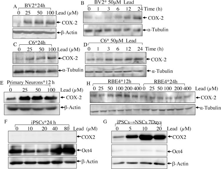
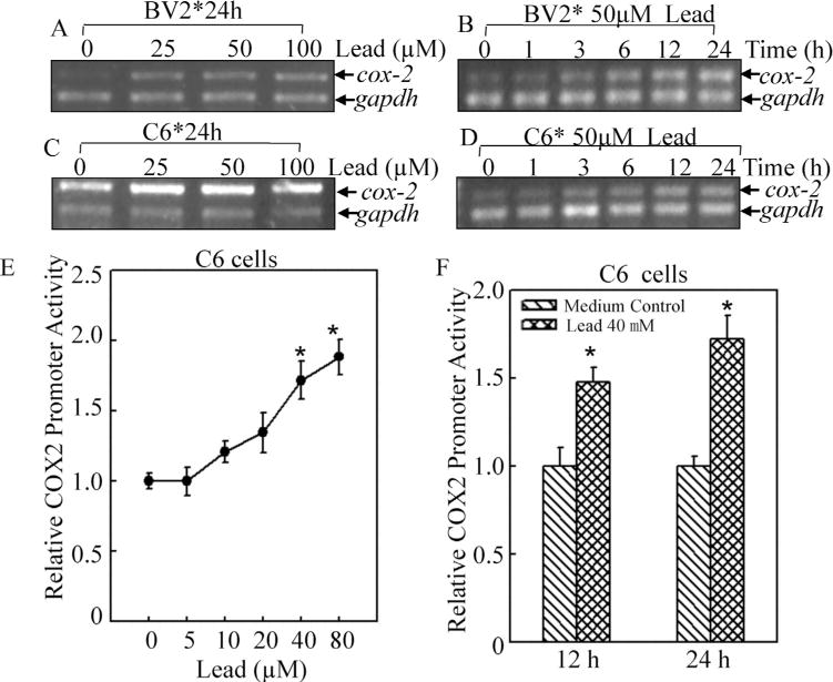
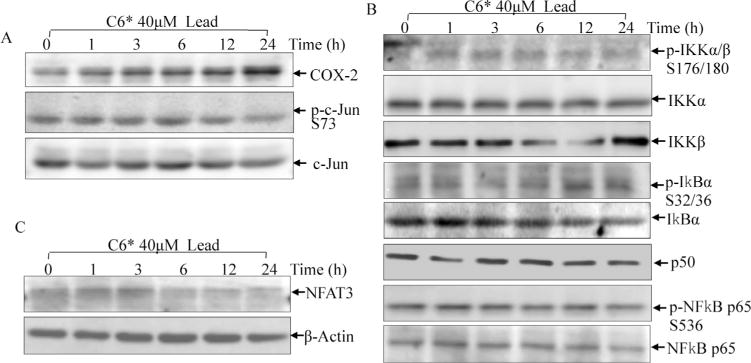
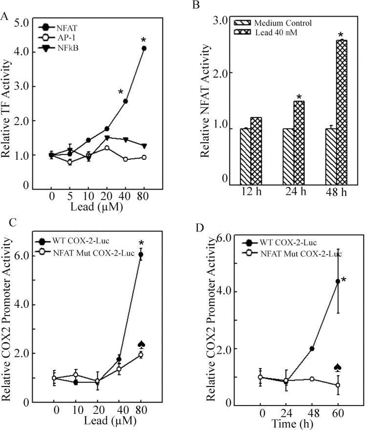
Epidemiologic studies have provided solid evidence for the neurotoxic effect of lead for decades of years. In view of the fact that children are more vulnerable to the neurotoxicity of lead, lead exposure has been an urgent public health concern. The modes of action of lead neurotoxic effects include disturbance of neurotransmitter storage and release, damage of mitochondria, as well as induction of apoptosis in neurons, cerebrovascular endothelial cells, astroglia and oligodendroglia. Our studies here, from a novel point of view, demonstrates that lead specifically caused induction of COX-2, a well known inflammatory mediator in neurons and glia cells. Furthermore, we revealed that COX-2 was induced by lead in a transcription-dependent manner, which relayed on transcription factor NFAT, rather than AP-1 and NFκB, in glial cells. Considering the important functions of COX-2 in mediation of inflammation reaction and oxidative stress, our studies here provide a mechanistic insight into the understanding of lead-associated inflammatory neurotoxicity effect via activation of pro-inflammatory NFAT3/COX-2 axis.
Keywords: COX-2; Lead; NFAT; Neurotoxicity.
Copyright © 2014 Elsevier Ireland Ltd. All rights reserved.
Conflict of interest statement
The authors declare that there are no conflicts of interest.
Figures
Similar articles
-
Extracellular HIV-Tat induces cyclooxygenase-2 in glial cells through activation of nuclear factor of activated T cells.J Immunol. 2008 Jan 1;180(1):530-40. doi: 10.4049/jimmunol.180.1.530. J Immunol. 2008. PMID: 18097055
-
Cyclooxygenase-2 induction by arsenite is through a nuclear factor of activated T-cell-dependent pathway and plays an antiapoptotic role in Beas-2B cells.J Biol Chem. 2006 Aug 25;281(34):24405-13. doi: 10.1074/jbc.M600751200. Epub 2006 Jun 29. J Biol Chem. 2006. PMID: 16809336
-
A cross-talk between NFAT and NF-κB pathways is crucial for nickel-induced COX-2 expression in Beas-2B cells.Curr Cancer Drug Targets. 2011 Jun;11(5):548-59. doi: 10.2174/156800911795656001. Curr Cancer Drug Targets. 2011. PMID: 21486220 Free PMC article.
-
Resveratrol modulates phorbol ester-induced pro-inflammatory signal transduction pathways in mouse skin in vivo: NF-kappaB and AP-1 as prime targets.Biochem Pharmacol. 2006 Nov 30;72(11):1506-15. doi: 10.1016/j.bcp.2006.08.005. Epub 2006 Sep 26. Biochem Pharmacol. 2006. PMID: 16999939 Review.
-
[Research progress on Pb-induced neurotoxicity through glial cells].Zhonghua Yu Fang Yi Xue Za Zhi. 2024 Oct 6;58(10):1610-1615. doi: 10.3760/cma.j.cn112150-20240513-00382. Zhonghua Yu Fang Yi Xue Za Zhi. 2024. PMID: 39428248 Review. Chinese.
Cited by
-
N,N'bis-(2-mercaptoethyl) isophthalamide (NBMI) exerts neuroprotection against lead-induced toxicity in U-87 MG cells.Arch Toxicol. 2021 Aug;95(8):2643-2657. doi: 10.1007/s00204-021-03103-2. Epub 2021 Jun 24. Arch Toxicol. 2021. PMID: 34165617
-
Alkaline phosphatase downregulation promotes lung adenocarcinoma metastasis via the c-Myc/RhoA axis.Cancer Cell Int. 2021 Apr 15;21(1):217. doi: 10.1186/s12935-021-01919-7. Cancer Cell Int. 2021. PMID: 33858415 Free PMC article.
-
Downregulation of miR-200c stabilizes XIAP mRNA and contributes to invasion and lung metastasis of bladder cancer.Cell Adh Migr. 2019 Dec;13(1):236-248. doi: 10.1080/19336918.2019.1633851. Cell Adh Migr. 2019. PMID: 31240993 Free PMC article.
-
Lead (Pb) as a Factor Initiating and Potentiating Inflammation in Human THP-1 Macrophages.Int J Mol Sci. 2020 Mar 24;21(6):2254. doi: 10.3390/ijms21062254. Int J Mol Sci. 2020. PMID: 32214022 Free PMC article.
-
How does NFAT3 regulate the occurrence of cardiac hypertrophy?Int J Cardiol Heart Vasc. 2023 Sep 20;48:101271. doi: 10.1016/j.ijcha.2023.101271. eCollection 2023 Oct. Int J Cardiol Heart Vasc. 2023. PMID: 37753338 Free PMC article. Review.
References
-
- Breder C, et al. Distribution and characterization of cyclooxygenase immunoreactivityin the ovine brain. J Comp Neurol. 1992;322(3):409–438. - PubMed
-
- Ding J, et al. Cyclooxygenase-2 induction by arsenite is through a nuclear factor of activated T-cell-dependent pathway and plays an antiapoptotic role in beas-2B cells. J Biol Chem. 2006;281(34):24405–24413. - PubMed
-
- Farkhondeh T, Boskabady M, Koohi M, Sadeghi-Hashjin G, Moin M. The effect oflead exposure on selected blood inflammatory biomarkers in guinea pigs. Cardiovasc. Hematol Disord Drug Targets. 2013;13(1):45–49. - PubMed
Publication types
MeSH terms
Substances
Grants and funding
LinkOut - more resources
Full Text Sources
Other Literature Sources
Molecular Biology Databases
Research Materials
Miscellaneous
