. 2014 Nov 1;74(21):6184-93.
doi: 10.1158/0008-5472.CAN-14-1357. Epub 2014 Sep 5.

Genetic deletion of AEG-1 prevents hepatocarcinogenesis

Affiliations

Genetic deletion of AEG-1 prevents hepatocarcinogenesis

Chadia L Robertson et al. Cancer Res. .

Abstract

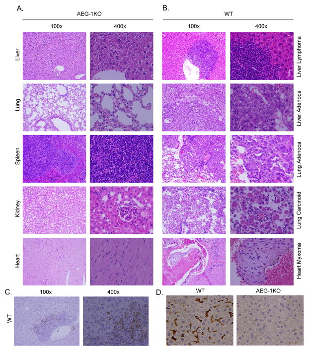
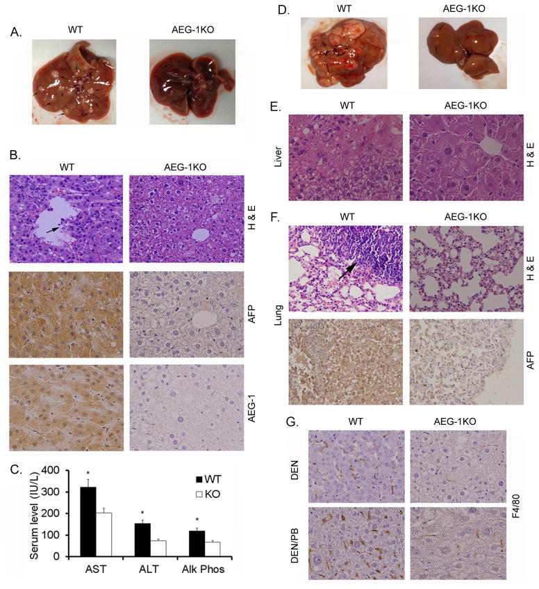
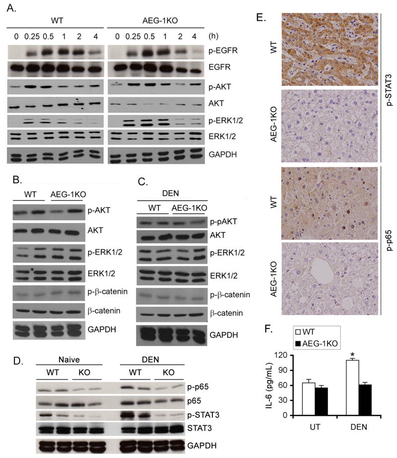
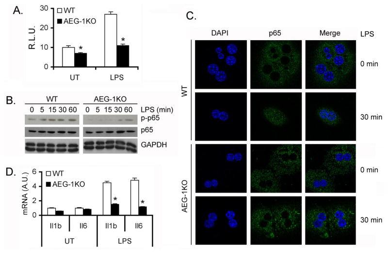
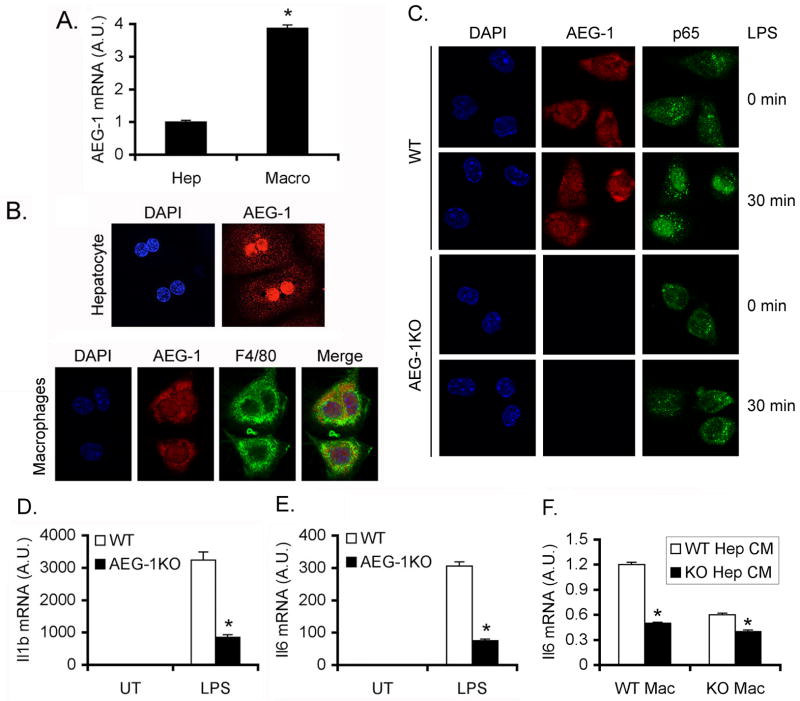
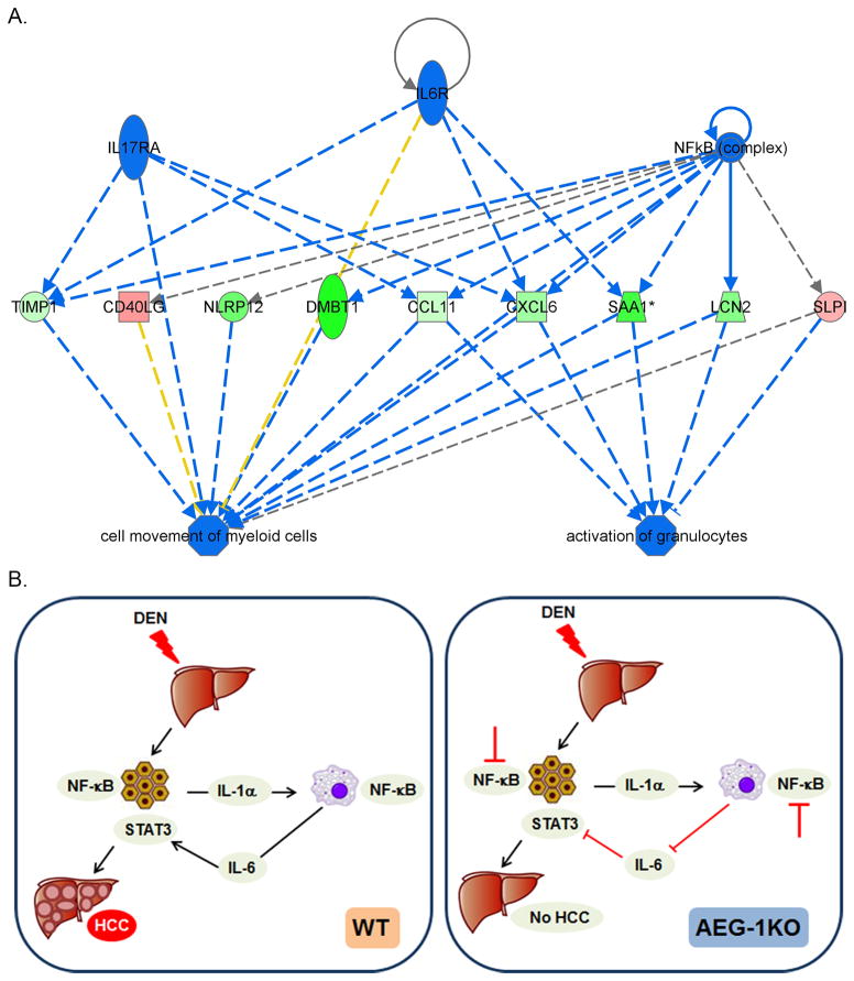
Activation of the oncogene AEG-1 (MTDH, LYRIC) has been implicated recently in the development of hepatocellular carcinoma (HCC). In mice, HCC can be initiated by exposure to the carcinogen DEN, which has been shown to rely upon activation of NF-κB in liver macrophages. Because AEG-1 is an essential component of NF-κB activation, we interrogated the susceptibility of mice lacking the AEG-1 gene to DEN-induced hepatocarcinogenesis. AEG-1-deficient mice displayed resistance to DEN-induced HCC and lung metastasis. No difference was observed in the response to growth factor signaling or activation of AKT, ERK, and β-catenin, compared with wild-type control animals. However, AEG-1-deficient hepatocytes and macrophages exhibited a relative defect in NF-κB activation. Mechanistic investigations showed that IL6 production and STAT3 activation, two key mediators of HCC development, were also deficient along with other biologic and epigenetics findings in the tumor microenvironment, confirming that AEG-1 supports an NF-κB-mediated inflammatory state that drives HCC development. Overall, our findings offer in vivo proofs that AEG-1 is essential for NF-κB activation and hepatocarcinogenesis, and they reveal new roles for AEG-1 in shaping the tumor microenvironment for HCC development.


Figures

Fig. 1
Fig. 1
AEG-1KO mice are resistant to age-associated spontaneous tumorigenesis. A. H & E staining of the indicated tissue sections of aged (16 m) AEG-1KO mouse demonstrating maintenance of normal histology and architecture. B. H & E staining of sections of liver, lung and heart of aged (16 m) WT mouse showing tumors. C. Immunohistochemical staining for AEG-1 in liver lymphoma of WT mouse. D. F4/80 staining for macrophages in liver sections of aged (16 m) WT and AEG-1KO mouse.
Fig. 2
Fig. 2
AEG-1KO mouse is resistant to experimental hepatocarcinogenesis. A. Representative photograph of the livers of WT and AEG-1KO mice 32 weeks after DEN injection. B, top panel, H & E staining of liver sections; middle panel, AFP staining of the liver sections; bottom panel, AEG-1 staining of the liver sections. These samples were collected at 32 weeks after DEN injection. C. Serum levels of the indicated liver enzymes at the end of the study. AST: aspartate aminotransferase, ALT; alanine aminotransferase and Alk Phos: alkaline phosphatase. Data represent mean ± SEM. n = 12 for WT and n = 8 for AEG-1KO. *: p<0.01. D. Representative photograph of the livers of WT and AEG-1KO mice 28 weeks after initial DEN injection receiving PB in drinking water. E. H & E staining of liver sections at 28 weeks. F. top panel, H & E staining of the lungs showing lung metastasis in WT mouse; bottom panel, AFP staining of a metastatic lung tumor in WT mouse. G. F4/80 staining for macrophages in liver sections of WT and AEG-1KO mice treated with DEN alone (top panels) or DEN/PB (bottom panels).
Fig. 3
Fig. 3
NF-κB and STAT3 activation is inhibited in AEG-1KO mouse. A. WT and AEG-1KO hepatocytes were treated with EGF (50 ng/mL) for the indicated time points and Western blotting was performed for the indicated proteins. B. Western blot was performed for the indicated proteins using liver lysates from adult WT and AEG-1KO mice. C. Western blotting was performed for the indicated proteins using liver lysates from DEN-treated WT and AEG-1KO mice at the end of the study. D. Western blotting was performed for the indicated proteins using liver lysates from naïve and DEN-treated WT and AEG-1KO mice at the end of the study. For B–D, each lane represents one independent mouse. E. DEN-treated liver sections were stained for p-STAT3 and p-p65 NF-κB at the end of the study. F. IL-6 protein level was measured in DEN-treated liver homogenates by ELISA. Data represent mean ± SEM. n = 5. *: p<0.01.
Fig. 4
Fig. 4
NF-κB activation is inhibited in AEG-1KO hepatocytes. A. NF-κB luciferase reporter activity was measured in WT and AEG-1KO hepatocytes. Firefly luciferase activity was normalized by Renilla luciferase activity. The activity of empty pGL3-basic vector was considered as 1. R.L.U.: Relative luciferase units. Data represent mean ± SEM of three independent experiments. *: p<0.01. B. WT and AEG-1KO hepatocytes were treated with LPS for the indicated time points and Western blotting was performed for the indicated proteins. C. Immunofluorescence followed by confocal microscopy of WT and AEG-1KO hepatocytes after LPS treatment for 30 min showing p65 nuclear translocation. D. WT and AEG-1KO hepatocytes were treated with LPS for 4 h and the mRNA level of Il1b and Il6 was measured by Taqman Q-RT-PCR. Data represent mean ± SEM of three independent experiments. *: p<0.01.
Fig. 5
Fig. 5
NF-κB activation is inhibited in AEG-1KO macrophages. A. AEG-1 mRNA level in hepatocytes and macrophages of WT mice measured by Taqman Q-RT-PCR. A.U.: arbitrary unit. Data represent mean ± SEM of three independent experiments. *: p<0.01. B. Immunofluorescence staining showing subcellular localization of AEG-1 in WT hepatocytes and macrophages. F4/80 staining was performed in macrophages to demonstrate authenticity of purification. C. Immunofluorescence followed by confocal microscopy of WT and AEG-1KO macrophages after LPS treatment for 30 min showing p65 nuclear translocation. D–E. WT and AEG-1KO macrophages were treated with LPS for 12 h and the mRNA levels of Il1b (D) and Il6 (E) were measured by Taqman Q-RT-PCR. F. WT and AEG-1KO macrophages were treated with conditioned media (CM) from LPS-treated WT and AEG-1KO hepatocytes and Il6 mRNA expression was measured by Taqman Q-RT-PCR 4 h later. For D–F, data represent mean ± SEM of three independent experiments. *: p<0.01.
Fig. 6
Fig. 6
Molecular mechanism rendering AEG-1KO mice resistant to HCC. A. Functional endpoints, analyzed by Ingenuity pathway analysis, that are inhibited in AEG-1KO mice. Blue line: inhibition; red line: activation; yellow line: finding inconsistent with state of downstream molecules; gray line: effect not predicted. Green color: downregulation of expression; red color: upregulation of expression. B. Schematic representation of the molecular mechanism of resistance of AEG-1KO mice to HCC. In WT mice DEN-induced hepatocyte injury leads to IL-1α production that activates NF-κB in the macrophages leading to production of IL-6. IL-6 activates STAT3 in hepatocytes facilitating proliferation of the mutated cells leading to HCC. In AEG-1KO mice NF-κB activation is inhibited in hepatocytes and macrophages thereby inhibiting IL-6 production and STAT3 activation. As a consequence, HCC development is significantly abrogated.

References

    1. El-Serag HB. Hepatocellular carcinoma. N Engl J Med. 2011;365:1118–27. - PubMed
    1. Berasain C, Castillo J, Perugorria MJ, Latasa MU, Prieto J, Avila MA. Inflammation and liver cancer: new molecular links. Ann N Y Acad Sci. 2009;1155:206–21. - PubMed
    1. Pikarsky E, Porat RM, Stein I, Abramovitch R, Amit S, Kasem S, et al. NF-kappaB functions as a tumour promoter in inflammation-associated cancer. Nature. 2004;431:461–6. - PubMed
    1. Karin M, Ben-Neriah Y. Phosphorylation meets ubiquitination: the control of NF-[kappa]B activity. Annu Rev Immunol. 2000;18:621–63. - PubMed
    1. Liu P, Kimmoun E, Legrand A, Sauvanet A, Degott C, Lardeux B, et al. Activation of NF-kappa B, AP-1 and STAT transcription factors is a frequent and early event in human hepatocellular carcinomas. J Hepatol. 2002;37:63–71. - PubMed

Publication types

MeSH terms