. 2014 Nov;184(11):3013-25.
doi: 10.1016/j.ajpath.2014.07.019. Epub 2014 Sep 2.

Pro-regenerative signaling after acetaminophen-induced acute liver injury in mice identified using a novel incremental dose model

Affiliations

Pro-regenerative signaling after acetaminophen-induced acute liver injury in mice identified using a novel incremental dose model

Bharat Bhushan et al. Am J Pathol. 2014 Nov.

Abstract

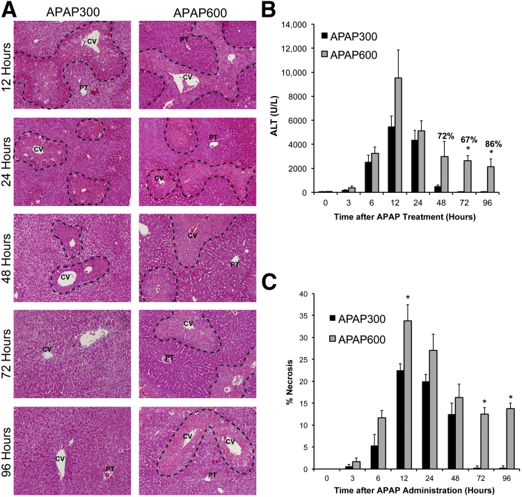
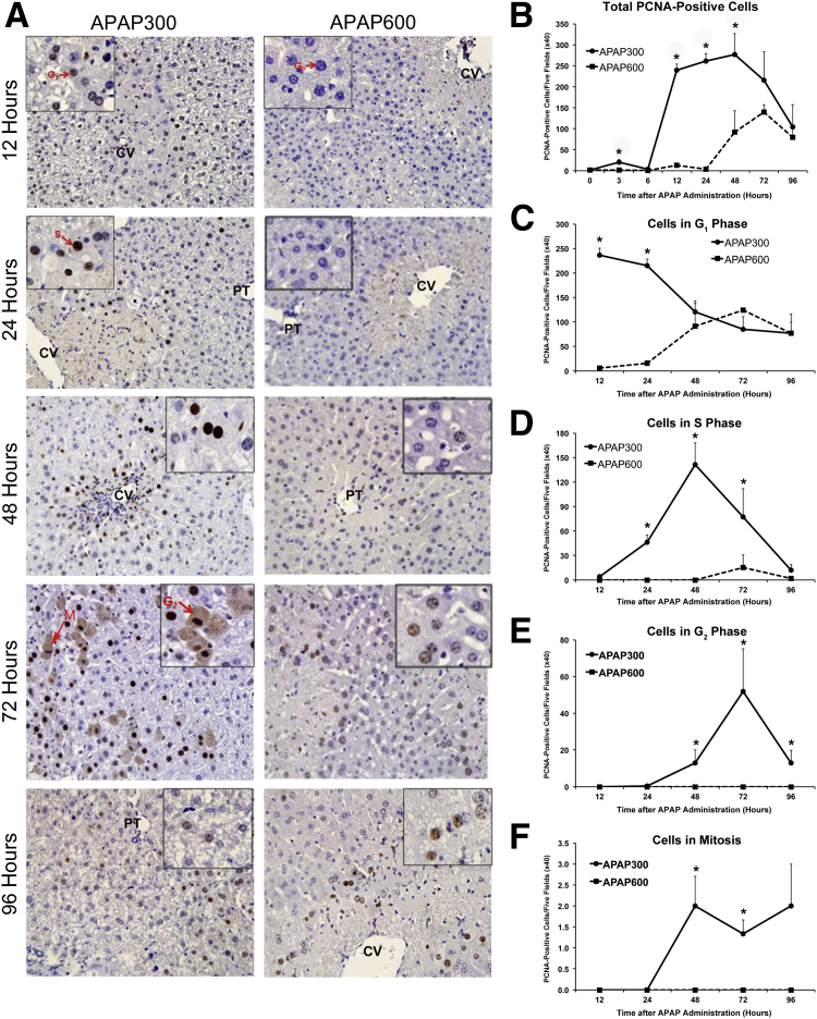
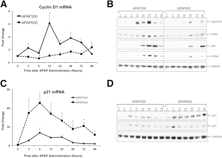
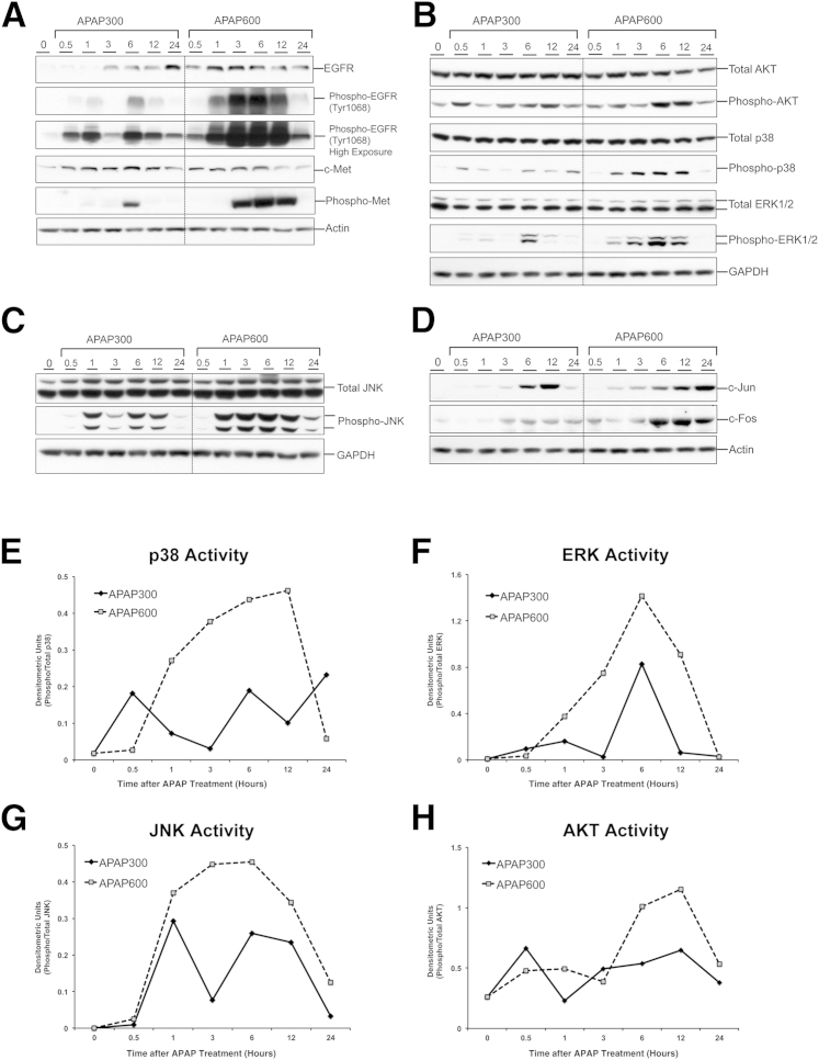
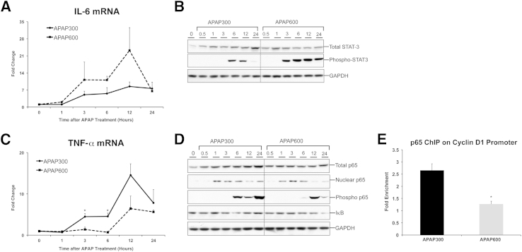
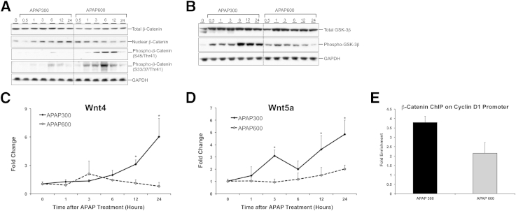
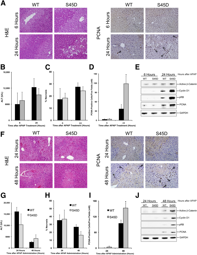
Acetaminophen (APAP) overdose results in acute liver failure and has limited treatment options. Previous studies show that stimulating liver regeneration is critical for survival after APAP overdose, but the mechanisms remain unclear. In this study, we identified major signaling pathways involved in liver regeneration after APAP-induced acute liver injury using a novel incremental dose model. Liver injury and regeneration were studied in C57BL/6 mice treated with either 300 mg/kg (APAP300) or 600 mg/kg (APAP600) APAP. Mice treated with APAP300 developed extensive liver injury and robust liver regeneration. In contrast, APAP600-treated mice exhibited significant liver injury but substantial inhibition of liver regeneration, resulting in sustained injury and decreased survival. The inhibition of liver regeneration in the APAP600 group was associated with cell cycle arrest and decreased cyclin D1 expression. Several known regenerative pathways, including the IL-6/STAT-3 and epidermal growth factor receptor/c-Met/mitogen-activated protein kinase pathways, were activated, even at APAP600, where regeneration was inhibited. However, canonical Wnt/β-catenin and NF-κB pathways were activated only in APAP300-treated mice, where liver regeneration was stimulated. Furthermore, overexpression of a stable form of β-catenin, where serine 45 is mutated to aspartic acid, in mice resulted in improved liver regeneration after APAP overdose. Taken together, our incremental dose model has identified a differential role of several signaling pathways in liver regeneration after APAP overdose and highlighted canonical Wnt signaling as a potential target for regenerative therapies for APAP-induced acute liver failure.


Figures

Figure 1
Figure 1
Sustained liver injury and inhibited recovery after higher dose of APAP. A: Representative photomicrographs of H&E-stained liver sections with necrotic area outlined. B: Bar graph shows serum ALT levels with percentage survival specified over bars at time points where any mortality was observed. C: Bar graph shows percentage necrosis area on the basis of H&E-stained liver sections. All samples were collected from mice treated with either APAP300 or APAP600 at various time points up to 96 hours after APAP treatment. P < 0.05 between two doses. CV, central vein; PT, portal triad.
Figure 2
Figure 2
Inhibition of liver regeneration following higher doses of APAP. A: Representative photomicrographs of PCNA-stained liver sections. Insets: Cells in specific phase of cell cycle (arrows), indicating different phases of the cell cycle. G0, G1, and S cells show blue, light brown, and deep brown nuclear staining, respectively; G2 cells, diffused brown cytoplasmic staining; and M phase cells, deep blue chromosomal staining. B: Line graph shows total number of PCNA-positive cells per five high-power (×40) fields. C–F: Line graphs show total number of cells in G1 (C), S (D), G2 (E), and M (F) phases per five high-power (×40) fields, demonstrating cell cycle progression. All samples were collected from mice treated with either APAP300 or APAP600 over a time course of 0 to 96 hours after APAP treatment. P < 0.05 between two doses. CV, central vein; PT, portal triad.
Figure 3
Figure 3
Cell cycle regulators correlate with inhibited liver regeneration at higher dose. Cyclin D1 (A) and p21 (C) mRNA expression, cyclin D1, CDK4, phospho-Rb, and PCNA (B) and p21 and p27 (D) protein expression in liver of mice treated with either APAP300 or APAP600. All samples were collected over a time course of 0 to 96 hours after APAP treatment. P < 0.05 between two doses. GAPDH, glyceraldehye-3-phosphate dehydrogenase.
Figure 4
Figure 4
Growth factor signaling via EGFR and c-Met and MAPK signaling remain highly activated after high dose of APAP. Western blot analysis of EGFR, phospho-EGFR (normal and high exposures), c-Met, phospho-Met (A), total AKT, phospho-AKT, total p38, phospho-p38, total ERK1/2, phospho-ERK 1/2 (B), total JNK, phospho-JNK (C), and c-Jun and c-Fos (D) using total liver extract of mice liver treated with either APAP300 or APAP600. Densitometric analysis shows p38 (E), ERK1/2 (F), JNK (G), and AKT (H) activation. All samples were collected over a time course of 0 to 24 hours after APAP treatment. GAPDH, glyceraldehye-3-phosphate dehydrogenase.
Figure 5
Figure 5
Differential activation of cytokine pathways during liver regeneration after APAP overdose. IL-6 (A) and TNF-α (C) mRNA expression in liver of mice treated with either APAP300 or APAP600. Western blot analysis of total STAT-3, phospho STAT-3 (B), total, nuclear, phosphorylated p65, and IκB (D) using total liver extract (unless specified) of mice treated with either APAP300 or APAP600. All samples were collected over a time course of 0 to 24 hours after APAP treatment. E: ChIP analysis shows p65 binding to cyclin D1 promoter at 12 hours after either APAP300 or APAP600 treatment. P < 0.05 between two doses. GAPDH, glyceraldehye-3-phosphate dehydrogenase.
Figure 6
Figure 6
Activation of Wnt/β-catenin signaling is inhibited after a higher dose of APAP. Western blot analysis of total β-catenin, nuclear β-catenin, phospho-β-catenin (S45/Thr41), phospho-β-catenin (S33/37/Thr41) (A) and total GSK-3β and phospho-GSK-3β (B). Total liver extracts (unless specified) of mice treated with either APAP300 or APAP600 were used for Western blot analyses. mRNA expression of Wnt4 (C) and Wnt5a (D) in liver of mice treated with either APAP300 or APAP600. All samples were collected over a time course of 0 to 24 hours after APAP treatment. E: ChIP analysis shows β-catenin binding to cyclin D1 promoter at 12 hours after either APAP300 or APAP600 treatment. P < 0.05 between two doses. GAPDH, glyceraldehye-3-phosphate dehydrogenase.
Figure 7
Figure 7
Overexpression of stable form of β-catenin improves liver regeneration after APAP overdose. A: Representative photomicrographs of H&E- and PCNA-stained liver sections from WT and S45D mice treated with 300 mg/kg APAP. B–D: Bar graphs show serum ALT levels (B), percentage necrosis area (C), and PCNA counts (D) in WT (black bars) and S45D (gray bars) mice treated with APAP300. E: Western blot analysis of active β-catenin, cyclin D1, pRb, and PCNA using total liver extracts in WT and S45D mice treated with APAP300. F: Representative photomicrographs of H&E- and PCNA-stained liver sections from WT and S45D mice treated with APAP600. Bar graphs show serum ALT levels (G), percentage necrosis area (H), and PCNA counts (I) in WT (black bars) and S45D (gray bars) mice treated with APAP600. J: Western blot analysis of active β-catenin, cyclin D1, pRb, and PCNA using total liver extracts in WT and S45D mice treated with APAP600. P < 0.05 between WT and S45D group. Arrowhead represents PCNA-positive cells. GAPDH, glyceraldehye-3-phosphate dehydrogenase.

References

    1. Nourjah P., Ahmad S.R., Karwoski C., Willy M. Estimates of acetaminophen (Paracetomal)-associated overdoses in the United States. Pharmacoepidemiol Drug Saf. 2006;15:398–405. - PubMed
    1. Athuraliya T.N., Jones A.L. Prolonged N-acetylcysteine therapy in late acetaminophen poisoning associated with acute liver failure: a need to be more cautious? Crit Care. 2009;13:144. - PMC - PubMed
    1. Keeffe E.B. Liver transplantation: current status and novel approaches to liver replacement. Gastroenterology. 2001;120:749–762. - PubMed
    1. Schmidt L.E., Dalhoff K. Alpha-fetoprotein is a predictor of outcome in acetaminophen-induced liver injury. Hepatology. 2005;41:26–31. - PubMed
    1. Apte U., Singh S., Zeng G., Cieply B., Virji M.A., Wu T., Monga S.P. Beta-catenin activation promotes liver regeneration after acetaminophen-induced injury. Am J Pathol. 2009;175:1056–1065. - PMC - PubMed

Publication types