. 2014 Oct 17;289(42):29420-36.
doi: 10.1074/jbc.M114.603068. Epub 2014 Sep 5.

Ring finger protein 34 (RNF34) interacts with and promotes γ-aminobutyric acid type-A receptor degradation via ubiquitination of the γ2 subunit

Affiliations

Ring finger protein 34 (RNF34) interacts with and promotes γ-aminobutyric acid type-A receptor degradation via ubiquitination of the γ2 subunit

Hongbing Jin et al. J Biol Chem. .

Abstract

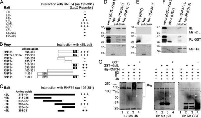
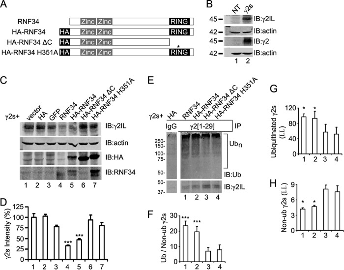
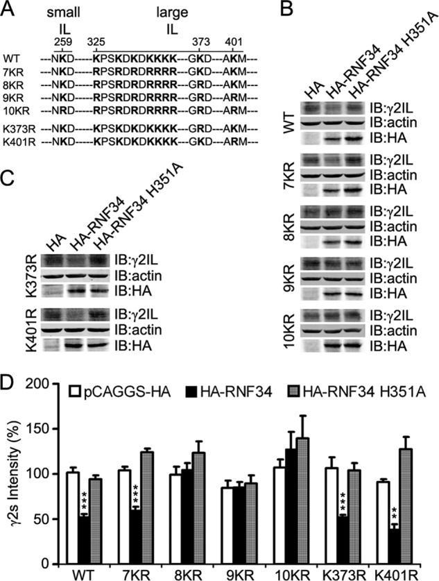
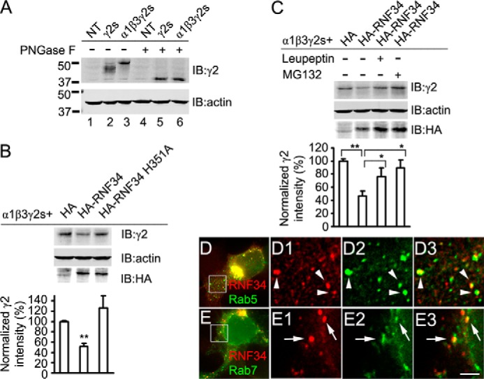
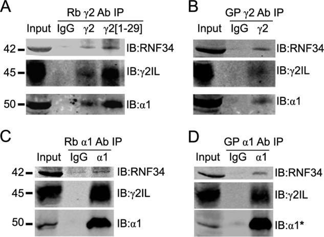
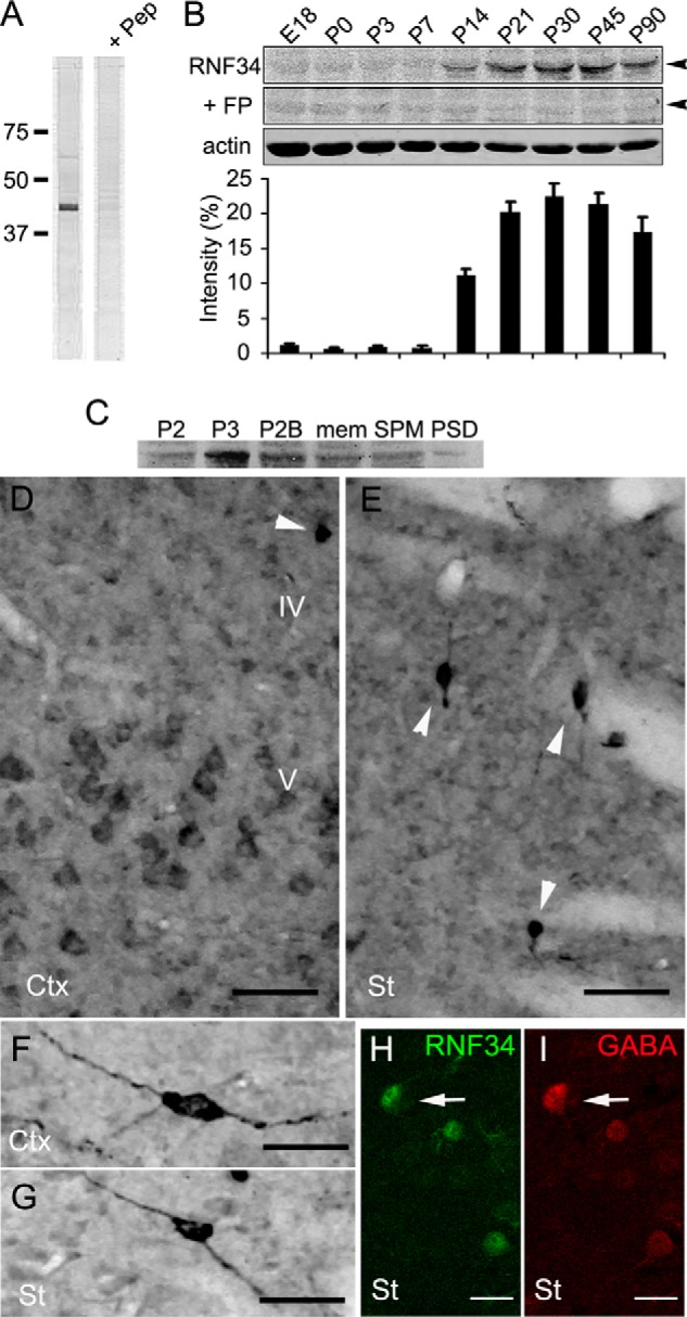
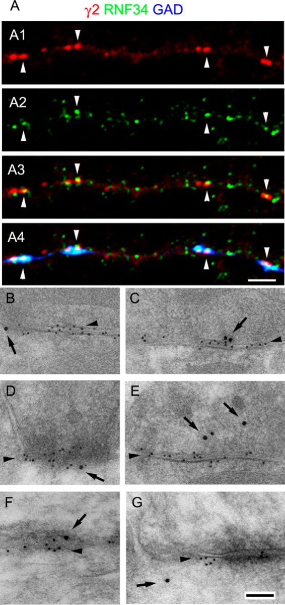
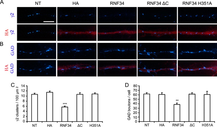
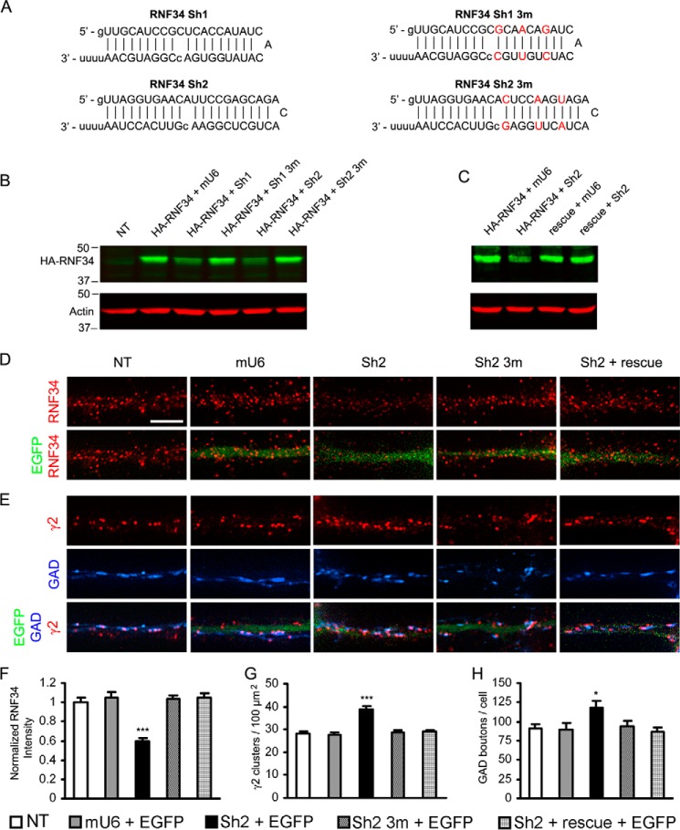
We have found that the large intracellular loop of the γ2 GABAA receptor (R) subunit (γ2IL) interacts with RNF34 (an E3 ubiquitin ligase), as shown by yeast two-hybrid and in vitro pulldown assays. In brain extracts, RNF34 co-immunoprecipitates with assembled GABAARs. In co-transfected HEK293 cells, RNF34 reduces the expression of the γ2 GABAAR subunit by increasing the ratio of ubiquitinated/nonubiquitinated γ2. Mutating several lysines of the γ2IL into arginines makes the γ2 subunit resistant to RNF34-induced degradation. RNF34 also reduces the expression of the γ2 subunit when α1 and β3 subunits are co-assembled with γ2. This effect is partially reversed by leupeptin or MG132, indicating that both the lysosomal and proteasomal degradation pathways are involved. Immunofluorescence of cultured hippocampal neurons shows that RNF34 forms clusters and that a subset of these clusters is associated with GABAergic synapses. This association is also observed in the intact rat brain by electron microscopy immunocytochemistry. RNF34 is not expressed until the 2nd postnatal week of rat brain development, being highly expressed in some interneurons. Overexpression of RNF34 in hippocampal neurons decreases the density of γ2 GABAAR clusters and the number of GABAergic contacts that these neurons receive. Knocking down endogenous RNF34 with shRNA leads to increased γ2 GABAAR cluster density and GABAergic innervation. The results indicate that RNF34 regulates postsynaptic γ2-GABAAR clustering and GABAergic synaptic innervation by interacting with and ubiquitinating the γ2-GABAAR subunit promoting GABAAR degradation.

Keywords: E3 Ubiquitin Ligase; GABAA Receptors; Interaction; Lysosome; Proteasome; RNF34; Synapse; Ubiquitination; Yeast Two-hybrid.


Figures

FIGURE 1.
FIGURE 1.
Characterization of the interaction between γ2IL and RNF34 and in vitro ubiquitination of γ2IL by RNF34. A–C, yeast two-hybrid. A, large intracellular loops (ILs) from various GABAA receptor subunits were used as bait to test for interaction with GS29, corresponding to RNF34 (aa 195–381). B, various fragments of RNF34 cDNA were tested as prey using γ2sIL as bait to determine the RNF34 domain that interacts with γ2sIL. ZZ and R represent the two zinc finger domains and the RING finger domain, respectively. The asterisk indicates the position of the H351A point mutation in the RING finger domain. C, various fragments of the γ2sIL were used as baits to map the domain that interacts with RNF34. D–F, in vitro pulldown of purified bacterial fusion proteins. D, GST-γ2sIL (35 kDa) was pulled down by immobilized His-RNF34-C (aa 195–381, 40 kDa) and His-RNF34-C* (aa 195–381, H351A, 40 kDa) but not by His (18 kDa). E, GST (27 kDa) was not pulled down by immobilized His-RNF34-C or His. F, GST-γ2sIL but not GST was pulled down by immobilized His-RNF34 FL (aa 1–381, 60 kDa). The GST-γ2sIL fusion protein reacted with both Ms anti-γ2IL and Rb anti-GST antibodies, whereas GST reacted with Rb anti-GST but not with Ms anti-γ2IL. His and His-tagged RNF34 fusion proteins were revealed with Ms anti-His. G, bacterial His-RNF34 fusion protein ubiquitinates GST-γ2sIL in an in vitro assay. Left immunoblot, smear corresponding to polyubiquitinated GST-γ2sIL (>55 kDa, Ubn) is observed when both His-RNF34 and GST-γ2sIL are present in the ubiquitination reaction (lane 3), as shown by immunoblotting (IB) with an anti-ubiquitin antibody (Ms Ub). No polyubiquitination smear is observed when GST is the substrate (lane 4). The smear is particularly strong at >100 kDa. Under the same reaction conditions, there is also an ∼55-kDa protein band (marked by *) corresponding to ubiquitinated GST-γ2sIL that reacted with Ms anti-Ub, Ms anti-γ2sIL, and Rb anti-GST in the three immunoblots (lane 3).
FIGURE 2.
FIGURE 2.
RNF34 is an E3 ligase that ubiquitinates γ2 subunit in transfected HEK293 cells. A, schematic representation of the RNF34 constructs. The position of the HA tag, zinc finger domains, and RING domain is indicated. The asterisk indicates the position of the H351A point mutation. B, expression of the γ2s subunit in HEK293 cells co-transfected with γ2s and pCAGGS-HA (γ2s, lane 2) was verified by immunoblotting (IB) with Ms anti-γ2IL and Rb anti-γ2 antibodies. Both antibodies recognize the ∼45-kDa γ2s protein band. Nontransfected cells (NT, lane 1) showed no expression of γ2s. C, RNF34, both nontagged (lane 4) and HA-tagged (lane 5), significantly reduced the expression of the γ2s subunit. HEK293 cells were co-transfected with γ2s, ubiquitin, and pCAGGS (vector, lane 1), pCAGGS-HA (HA, lane 2), or pCAGGS-GFP (GFP, lane 3), or a RNF34 construct (RNF34, HA-RNF34, HA-RNF34 ΔC, or HA-RNF34 H351A, lanes 4–7, respectively). RIPA extracts were subjected to immunoblotting with Ms anti-γ2IL, Ms anti-actin, Ms anti-HA, or Rb anti-RNF34 antibodies. The expression of RNF34 constructs was verified by HA immunoreactivity (lanes 5–7) and Rb anti-RNF34 antibody to the C terminus (lanes 4–7). Note that Rb anti-RNF34 recognizes a C terminus epitope and does not recognize HA-RNF34 ΔC (lane 6). HA-RNF34 migrated slightly slower than RNF34 as shown by immunoblotting with Rb anti-RNF34. Actin was used as the loading control. The expected HA control protein band (∼1.5 kDa) corresponding to pCAGGS-HA (lane 2) ran out of the gel. D, quantification of the γ2s subunit protein band from the various lanes in C normalized for actin and vector transfection (100%). Data are presented as mean ± S.E., n = 4 independent experiments. ***, p < 0.001 in one-way ANOVA Tukey-Kramer multiple comparison test. E and F, RNF34 and HA-RNF34 significantly increase the ratio of ubiquitinated/nonubiquitinated γ2s subunits. RIPA extracts of HEK293 cells, which were co-transfected with γ2s, Ub, and RNF34 constructs, were immunoprecipitated with Rb anti-γ2(1–29) antibody or nonimmune serum (IgG), and the precipitates were subjected to SDS-PAGE and immunoblotting with Ms anti-ubiquitin (Ub) or Ms anti-γ2IL mAbs. The bar graph below F shows the quantification of the ratio of the ubiquitination smear fluorescence intensity (>55 kDa, Ubn) to the 45-kDa nonubiquitinated γ2s subunit fluorescence intensity. The lane corresponding to the nonimmune serum (IgG) is a nonadjacent lane from the same Western blot as the other lanes 1–4. G, quantification of the integrated intensity (I.I.) of the smear of ubiquitinated γ2s shown in E. H, quantification of the integrated intensity (I.I.) of the nonubiquitinated 45 kDa γ2s of E. Data in F–H are presented as mean ± S.E., n = 3 independent experiments, *, p < 0.05 and ***, p < 0.001 in one-way ANOVA Tukey-Kramer multiple comparison test.
FIGURE 3.
FIGURE 3.
Combination of several lysine-to-arginine mutations in the IL makes the γ2s subunit resistant to the degradation by RNF34. A, illustration of the position of the lysine residue at the small intracellular loop (small IL, aa 259) and of the 9 lysine residues at the large intracellular loop (large IL, aa 325, 328, 330, 332, 333, 334, 335, 373, and 401) of the γ2s subunit that were mutated into arginines. The small IL (aa 258–260) is located between TM1 and TM2, and the large IL (aa 313–404) is between TM3 and TM4. B, HA-RNF34 significantly reduces the expression level of γ2s WT or 7KR, but it does not significantly affect the expression level of γ2s 8KR, 9KR, or 10KR, compared with the HA or HA-RNF34 H351A controls. HEK293 cells, co-transfected with a γ2s subunit (WT, 7KR, 8KR, 9KR, or 10KR), ubiquitin, and a RNF34 construct (HA-RNF34 or HA-RNF34 H351A) or pCAGGS-HA as control vector were lysed, subjected to SDS-PAGE, and immunoblotted (IB) with Ms anti-γ2IL, Ms anti-actin, or Ms anti-HA. C, expression level of γ2s K373R or K401R point mutants (the Lys-Arg point mutation of the 8th or 9th lysine residues in the large IL of γ2s subunit) was significantly reduced by HA-RNF34, but not by HA-RNF34 H351A. D, quantification of the γ2s subunit protein band intensity normalized for that of actin and pCAGGS-HA transfection control for WT. Data are presented as mean ± S.E., n = 4 independent experiments. **, p < 0.01; ***, p < 0.001, for each mutant, statistical significance was determined in one-way ANOVA Tukey-Kramer multiple comparison test.
FIGURE 4.
FIGURE 4.
RNF34 induces the degradation of α1β3γ2s GABAARs via the lysosome and proteasome pathways. A, γ2s subunit, when in HEK293 cells, is co-expressed with α1 and β3 subunits (α1β3γ2s, lane 3), has slower mobility than when γ2s is expressed alone (γ2s, lane 2), as shown by immunoblotting with Rb anti-γ2 antibody. NT represents nontransfected HEK293 cells. After digestion with peptide-N-glycosidase F (PNGase F), γ2s showed faster mobility that was identical in cells transfected with γ2s alone or with α1β3γ2s (lanes 5 and 6, respectively). B, HA-RNF34, but not HA-RNF34 H351A, significantly reduces the expression levels of 50-kDa γ2s in the α1β3γ2s recombinant receptors. Data are presented as mean ± S.E., n = 8 independent experiments; **, p < 0.01 in one-way ANOVA Tukey-Kramer multiple comparison test. C, leupeptin or MG132 partially reverse the effect of HA-RNF34 on the expression of the recombinant γ2s. HEK293 cells were co-transfected with α1, β3, γ2s subunit, ubiquitin, and a control vector pCAGGS-HA (HA) or HA-RNF34. After 3 days, cells were lysed and immunoblotted with Rb anti-γ2, Ms anti-actin, and Ms anti-HA antibodies. To the indicated cultures (+), leupeptin or MG132 was added 12 h before harvesting. The bar graphs below B and C show the expression level of γ2 subunits (fluorescence intensity) normalized for that of actin and that of the HA control. Data are presented as mean ± S.E., n = 8 independent experiments; *, p < 0.05; **, p < 0.01 in one-way ANOVA Tukey-Kramer multiple comparison test. D, HEK293 cells were co-transfected with RNF34 and the early endosome marker EGFP-Rab5, followed by immunofluorescence with anti-RNF34 and EGFP fluorescence. D1–D3, high magnification images of the boxed area in D. RNF34 (D1, red) frequently co-localizes with EGFP-Rab5 (D2, green) as shown in the overlay (D3, arrowheads). E, HEK293 cells were co-transfected with RNF34 and the late endosome marker EGFP-Rab7. E1–E3, high magnification images of the boxed area in E. RNF34 (E1, red) frequently co-localizes with EGFP-Rab7 (E2, green) as shown in the overlay (E3, arrows). Scale bar, 10 μm (D and E), 2.5 μm (D1–D3 and E1–E3).
FIGURE 5.
FIGURE 5.
RNF34 co-immunoprecipitates with brain GABAARs. A and B, various antisera to the γ2 GABAAR subunit, two raised in rabbit (Rb γ2 and Rb γ2(1–29), in A) and one in guinea pig (GP γ2, in B) co-precipitate RNF34 (42 kDa) with GABAARs from rat forebrain membrane extracts. GABAARs in the precipitate were revealed by the presence of γ2 (45 kDa) and α1 (50 kDa) GABAAR subunits. C and D, antisera to the α1 GABAAR subunit raised in rabbit (Rb α1, in C) or guinea pig (GP α1, in D) co-precipitate RNF34 and GABAARs, as shown by the presence of RNF34, γ2, and α1 subunits in the precipitate. In all panels, the presence of RNF34 and GABAARs in the precipitate was detected by immunoblotting with Rb anti-RNF34, Ms anti-γ2IL, Ms anti-α1, with the exception of α1 in D where Rb anti-α1* was used.
FIGURE 6.
FIGURE 6.
Expression and distribution of RNF34 in the rat brain. A, immunoblot with the Rb anti-RNF34 antibody of the rat forebrain crude synaptosomal (P2) fraction, shows a major protein band ∼42 kDa, which is displaced by 100 μg/ml of the antigenic peptide (+ Pep). 20 μg of the total proteins were loaded into each lane. B, immunoblots of rat forebrain homogenates of various ages from embryonic day 18 (E18) to postnatal day 90 (P90) with Rb anti-RNF34 and Ms anti-actin antibodies. 75 μg of total protein were loaded into each lane. The RNF34 antibody recognizes a 42-kDa protein band (arrowhead), whose immunoreactivity is displaced by incubating the antibody with 100 μg/ml of the purified bacterial His-RNF34 (aa 195–381) fusion protein (+ FP). The bar graph shows the quantification of the relative RNF34 protein expression levels by densitometry of the RNF34 protein band normalized to actin protein band density. C, immunoblot of various rat forebrain subcellular fractions with Rb anti-RNF34 antibody. The RNF34 protein band is present in all the fractions tested, including crude synaptosomal (P2), microsomal (P3), synaptosomal (P2B), total membrane (mem), SPM, and one Triton PSD fractions. 25 μg of the total proteins were loaded into each lane. D and E, RNF34 immunocytochemistry in layers IV and V of the cerebral cortex (Ctx) and corpus striatum (St). Arrowheads indicate neurons with high RNF34 immunoreactivity. F and G, individual neurons with high level of RNF34 immunoreactivity in the cerebral cortex and striatum, respectively. H and I, double label fluorescence confocal microscopy of corpus striatum from rat brain sections immunolabeled with anti-RNF34 (green in H) and anti-GABA (red in I). GABA+ interneurons show a high expression level of RNF34 protein (arrow). Scale bar, 50 μm (D and E), 25 μm (F and G), and 20 μm (H and I).
FIGURE 7.
FIGURE 7.
GABAergic synapses frequently have RNF34 associated with them. A, triple-labeled immunofluorescence of 21 DIV cultured hippocampal neurons with GP anti-γ2 GABAAR subunit (red, A1), Rb anti-RNF34 (green, A2), and sheep anti-GAD (blue) antibodies. A3 shows the overlay of γ2 and RNF34 fluorescence, whereas A4 shows the overlay of the three fluorescence channels. Arrowheads indicate GABAergic synapses (γ2+ and GAD+) that have co-localizing RNF34 clusters. Scale bar, 2.5 μm (A1–A4). B–G, postembedding EM immunogold double-labeling of rat brain cerebellum (B, C, and E) and cerebral cortex (D, F, and G) with Rb anti-RNF34 and mouse anti-β2/3 GABAAR subunits. The β2/3 immunolabeling (smaller gold particles) is indicated by arrowheads. GABAergic synapses are identified by their morphology and the concentration of β2/3 GABAAR subunits. The RNF34 immunolabeling (larger gold particles, arrows) is localized at or near GABAergic synapses (B–G), GABAergic presynaptic terminals (C, E, and F), and GABAergic postsynapses (B, D, and G). The goat anti-rabbit IgG and goat anti-mouse IgG secondary antibodies were conjugated to 18 and 10 nm diameter colloidal gold particles, respectively. Scale bar, 100 nm (B–G).
FIGURE 8.
FIGURE 8.
Cultured hippocampal neurons overexpressing RNF34 show reduced density of endogenous γ2-GABAAR clusters and reduced GABAergic innervations. A and B, representative immunofluorescence images of dendrites from hippocampal neurons that were nontransfected (NT) or transfected with EGFP and vector control (HA) or EGFP and various RNF34 constructs (HA-RNF34, HA-RNF34 ΔC, or HA-RNF34 H351A, all in pCAGGS plasmid). Primary antibodies were Ms anti-HA (red) in A and B, Rb anti-γ2 (blue in A), or sheep anti-GAD (blue in B). Neurons transfected with the RNF34 constructs or HA were identified by anti-HA fluorescence (red), which coincided with the fluorescence of the co-transfected EGFP (not shown for economy of space). The HA-RNF34 constructs were made in pCAGGS plasmid, and overexpression results were determined 2 days after transfection (13 DIV). Scale bar, 5 μm. C and D, quantification of the effect of RNF34 constructs on the density of γ2 clusters and the number of GAD boutons innervating the transfected neurons. Data are presented as mean ± S.E., **, p < 0.01; ***, p < 0.001 in one-way ANOVA Tukey-Kramer multiple comparison test, n = 16 neurons for each group from four independent transfection experiments.
FIGURE 9.
FIGURE 9.
Knocking down endogenous RNF34 in cultured hippocampal neurons increases both the γ2-GABAAR cluster density and GABAergic innervation. A, RNF34 shRNAs (Sh1 and Sh2) used in this study. The three point mutations in Sh1 3m and Sh2 3m are shown in red. B, Sh1 and Sh2 significantly knocked down the protein expression of HA-RNF34 (in pRK5 plasmid), compared with HEK293 cells co-transfected with HA-RNF34 and various control plasmids (mU6, Sh1 3m, or Sh2 3m). Cell lysates were immunoblotted with Ms anti-HA and Ms anti-actin mAbs. C, Sh2 did not knock down the protein expression of the rescue mRNA, although Sh2 knocked down the protein expression of the HA-RNF34. D and E, representative immunofluorescence images of dendrites from hippocampal neurons that were nontransfected (NT) or co-transfected with EGFP and mU6 pro vector (mU6), or various Sh2 constructs (Sh2 or Sh2 3m), or Sh2 + rescue. Immunofluorescence with Rb anti-RNF34 (red in D), Rb anti-γ2 (red in E) or sheep anti-GAD (blue in E). Dendrites of transfected neurons were identified by EGFP fluorescence (green). Scale bar, 5 μm. F–H, quantification of the effect of knocking down RNF34 with Sh2 on the RNF34 fluorescence intensity (F), γ2-GABAAR cluster density (G), and GAD boutons contacting the transfected neurons (H). *, p < 0.05; ***, p < 0.001 in one-way ANOVA Tukey-Kramer multiple comparison test, n = 16 neurons for each group from four independent transfection experiments.

Similar articles

Cited by

References

    1. Hershko A., Ciechanover A. (1998) The ubiquitin system. Annu. Rev. Biochem. 67, 425–479 - PubMed
    1. Hoppe T. (2005) Multiubiquitylation by E4 enzymes: “one size” doesn't fit all. Trends Biochem. Sci. 30, 183–187 - PubMed
    1. Lovering R., Hanson I. M., Borden K. L., Martin S., O'Reilly N. J., Evan G. I., Rahman D., Pappin D. J., Trowsdale J., Freemont P. S. (1993) Identification and preliminary characterization of a protein motif related to the zinc fingeret. Proc. Natl. Acad. Sci. U.S.A. 90, 2112–2116 - PMC - PubMed
    1. Pickart C. M. (2001) Mechanisms underlying ubiquitination. Annu. Rev. Biochem. 70, 503–533 - PubMed
    1. Deshaies R. J., Joazeiro C. (2009) RING domain E3 ubiquitin ligases. Annu. Rev. Biochem. 78, 399–434 - PubMed

Publication types

MeSH terms