Disruption of tumor suppressor gene Hint1 leads to remodeling of the lipid metabolic phenotype of mouse liver
- PMID: 25193995
- PMCID: PMC4617133
- DOI: 10.1194/jlr.M050682
Disruption of tumor suppressor gene Hint1 leads to remodeling of the lipid metabolic phenotype of mouse liver
Abstract
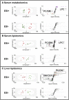
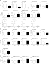
A lipidomic and metabolomic investigation of serum and liver from mice was performed to gain insight into the tumor suppressor gene Hint1. A major reprogramming of lipid homeostasis was found in both serum and liver of Hint1-null (Hint(-/-)) mice, with significant changes in the levels of many lipid molecules, as compared with gender-, age-, and strain-matched WT mice. In the Hint1(-/-) mice, serum total and esterified cholesterol were reduced 2.5-fold, and lysophosphatidylcholines (LPCs) and lysophosphatidic acids were 10-fold elevated in serum, with a corresponding fall in phosphatidylcholines (PCs). In the liver, MUFAs and PUFAs, including arachidonic acid (AA) and its metabolic precursors, were also raised, as was mRNA encoding enzymes involved in AA de novo synthesis. There was also a significant 50% increase in hepatic macrophages in the Hint1(-/-) mice. Several hepatic ceramides and acylcarnitines were decreased in the livers of Hint1(-/-) mice. The changes in serum LPCs and PCs were neither related to hepatic phospholipase A2 activity nor to mRNAs encoding lysophosphatidylcholine acetyltransferases 1-4. The lipidomic phenotype of the Hint1(-/-) mouse revealed decreased inflammatory eicosanoids with elevated proliferative mediators that, combined with decreased ceramide apoptosis signaling molecules, may contribute to the tumor suppressor activity of Hint1.
Keywords: apoptosis; carcinogenesis; cholesterol; eicosanoids; lipidomics; mass spectrometry; metabolomics; phospholipids; proliferation.
Copyright © 2014 by the American Society for Biochemistry and Molecular Biology, Inc.
Figures


Similar articles
-
Ablation of the tumor suppressor histidine triad nucleotide binding protein 1 is protective against hepatic ischemia/reperfusion injury.Hepatology. 2011 Jan;53(1):243-52. doi: 10.1002/hep.23978. Epub 2010 Dec 7. Hepatology. 2011. PMID: 21140474
-
Hint1 is a haplo-insufficient tumor suppressor in mice.Oncogene. 2006 Feb 2;25(5):713-21. doi: 10.1038/sj.onc.1209111. Oncogene. 2006. PMID: 16186798
-
Hepatic lipid profile in mice fed a choline-deficient, low-methionine diet resembles human non-alcoholic fatty liver disease.Lipids Health Dis. 2020 Dec 9;19(1):250. doi: 10.1186/s12944-020-01425-1. Lipids Health Dis. 2020. PMID: 33298075 Free PMC article.
-
[HINT1--a novel tumor suppressor protein of the HIT superfamily].Postepy Biochem. 2010;56(1):55-60. Postepy Biochem. 2010. PMID: 20499681 Review. Polish.
-
Emerging Roles of Histidine Triad Nucleotide Binding Protein 1 in Neuropsychiatric Diseases.Zhongguo Yi Xue Ke Xue Yuan Xue Bao. 2017 Oct 30;39(5):705-714. doi: 10.3881/j.issn.1000-503X.2017.05.018. Zhongguo Yi Xue Ke Xue Yuan Xue Bao. 2017. PMID: 29125116 Review. English.
Cited by
-
VANGL2 downregulates HINT1 to inhibit the ATM-p53 pathway and promote cisplatin resistance in small cell lung cancer.Cell Death Discov. 2025 Apr 8;11(1):153. doi: 10.1038/s41420-025-02424-w. Cell Death Discov. 2025. PMID: 40199845 Free PMC article.
-
Circulating MicroRNA-122 Is Associated With the Risk of New-Onset Metabolic Syndrome and Type 2 Diabetes.Diabetes. 2017 Feb;66(2):347-357. doi: 10.2337/db16-0731. Epub 2016 Nov 29. Diabetes. 2017. PMID: 27899485 Free PMC article.
-
Axonal neuropathy with neuromyotonia: there is a HINT.Brain. 2017 Apr 1;140(4):868-877. doi: 10.1093/brain/aww301. Brain. 2017. PMID: 28007994 Free PMC article. Review.
-
Hepatitis C Virus Infection Upregulates Plasma Phosphosphingolipids and Endocannabinoids and Downregulates Lysophosphoinositols.Int J Mol Sci. 2023 Jan 11;24(2):1407. doi: 10.3390/ijms24021407. Int J Mol Sci. 2023. PMID: 36674922 Free PMC article.
-
The plasma lipidome in acute myeloid leukemia at diagnosis in relation to clinical disease features.BBA Clin. 2017 Mar 8;7:105-114. doi: 10.1016/j.bbacli.2017.03.002. eCollection 2017 Jun. BBA Clin. 2017. PMID: 28331812 Free PMC article.
References
-
- Fujiwara M., Marusawa H., Wang H. Q., Iwai A., Ikeuchi K., Imai Y., Kataoka A., Nukina N., Takahashi R., Chiba T. 2008. Parkin as a tumor suppressor gene for hepatocellular carcinoma. Oncogene. 27: 6002–6011. - PubMed
Publication types
MeSH terms
Substances
Grants and funding
LinkOut - more resources
Full Text Sources
Other Literature Sources
Molecular Biology Databases

