. 2014 Nov 15;592(22):5109-27.
doi: 10.1113/jphysiol.2014.274498. Epub 2014 Sep 5.

Bradykinin enhances invasion of malignant glioma into the brain parenchyma by inducing cells to undergo amoeboid migration

Affiliations

Bradykinin enhances invasion of malignant glioma into the brain parenchyma by inducing cells to undergo amoeboid migration

Stefanie Seifert et al. J Physiol. .

Abstract

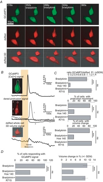
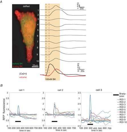
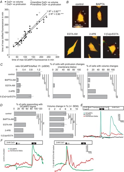
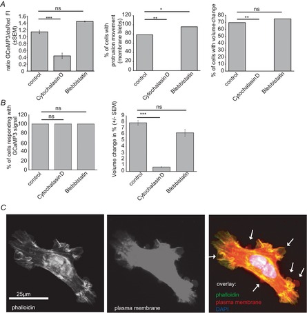
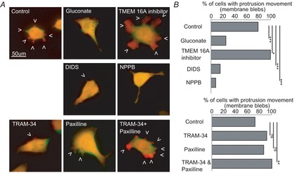
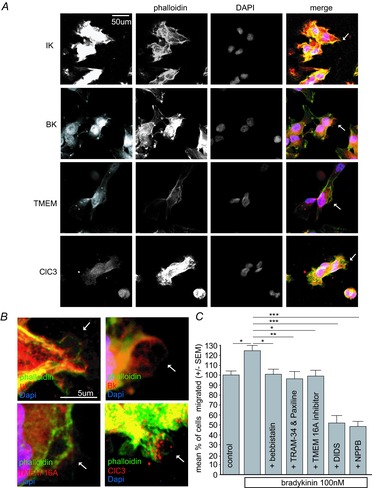
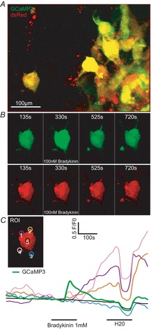
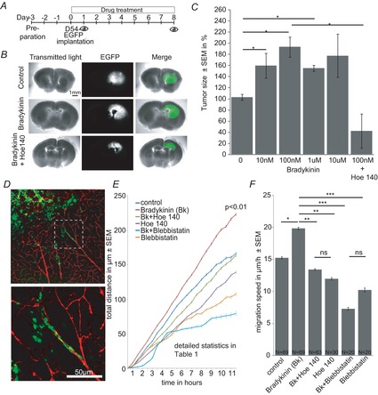
The molecular and cellular mechanisms governing cell motility and directed migration in response to the neuropeptide bradykinin are largely unknown. Here, we demonstrate that human glioma cells whose migration is guided by bradykinin generate bleb-like protrusions. We found that activation of the B2 receptor leads to a rise in free Ca(2+) from internal stores that activates actomyosin contraction and subsequent cytoplasmic flow into protrusions forming membrane blebs. Furthermore Ca(2+) activates Ca(2+)-dependent K(+) and Cl(-) channels, which participate in bleb regulation. Treatment of gliomas with bradykinin in situ increased glioma growth by increasing the speed of cell migration at the periphery of the tumour mass. To test if bleb formation is related to bradykinin-promoted glioma invasion we blocked glioma migration with blebbistatin, a blocker of myosin kinase II, which is necessary for proper bleb retraction. Our findings suggest a pivotal role of bradykinin during glioma invasion by stimulating amoeboid migration of glioma cells.


Figures

Figure 1
Figure 1. Bradykinin induces an increase of intracellular [Ca2+] via B2 receptor which is accompanied by protrusion and volume changes
A, confocal time lapse series of D54 glioma cells in vitro before, during and after bradykinin application (100 nm, 120 s, 37°C). GCaMP3 fluorescence (upper series) and dsRed fluorescence (middle series) were recorded in parallel. GCaMP3 fluorescence was recorded in one confocal z-plane, whereas dsRed fluorescence was recorded in 14 confocal planes (1 μm intervals). We used dsRed fluorescence to determine protrusion movement by analysing z-projections. Furthermore we used dsRed fluorescence for calculating the cell's volume by 3D reconstruction for every time point (lowest series). B, quantification of the data shown in A. Upper trace shows GCaMP3 fluorescence ([Ca2+]i), middle trace dsRed fluorescence (protrusion movement) and lowest trace volume recordings during bradykinin application (indicated by the yellow box). White circles in the inserts indicate the regions of interest (ROI) that were analysed. C, summary bar graphs of GCaMP3 fluorescence intensity (FI), percentage of cells with protrusion movement and percentage of cells showing volume changes. D, summary bar graphs of percentage of cells responding with a GCaMP3 signal and percentage volume changes when treated with 100 nm bradykinin (n = 27), 100 nm bradykinin + 5 μm Hoe-140 (n = 18) or 100 nm bradykinin + 5 μm R715 (n = 18). One-way-ANOVA with a Bonferroni post hoc test was used to compare GCaMP3 FI/dsRed and volume changes (%). χ2 test was used to compare the numbers of cells.
Figure 2
Figure 2. Analysis of glioma cell movement during bradykinin application
A, dsRed fluorescence of z-projections (see also Supporting information Video S1) for every time point was analysed within ROI 1–10 (indicated by dashed white circles). Picture shows cell before (green) and during a 2 min 100 nm bradykinin application (red). On the right side individual traces of every ROI are shown. Bradykinin application is indicated by a yellow box. For comparison traces of [Ca2+]i and cell volume are shown at the bottom. Notice that D54 cells show shape changes only in certain compartments of the cell. Membrane blebs occurred mainly in the periphery. B, dsRed fluorescence of 3 example glioma cells analysed like the cell in A. Traces have been normalized to mean baseline intensity (F0) Black bar indicates application of 100 nm bradykinin for 2 min.
Figure 3
Figure 3. Ca2+ dependence of membrane blebbing and volume changes
A, temporal Pearson-Correlation analysis of the maximal [Ca2+]i compared with the maximum of protrusion movement or maximum of volume change. B–D, D54 cells were incubated with blockers of the Ca2+ pathway before bradykinin application. Controls received 100 nm bradykinin for 2 min without pretreatment. B, pictures show dsRed fluorescence of example cells before (green) and during (red) 100 nm bradykinin application (overlay yellow). C, summary bar graphs of GCaMP3 fluorescence intensity (FI), percentage of cells with protrusion movement and percentage of cells showing volume changes. D, summary bar graphs of percentage of cells responding with a GCaMP3 signal and volume changes (%). Controls n = 23, 5 μm BAPTA-AM n = 22, 5 μm EGTA-AM n = 15, 100 μm 2-APB n = 29, 0[Ca2+]o/1 mm EGTA n = 34. One-way-ANOVA with a Bonferroni post hoc testwas used to compare GCaMP3 FI/dsRed and volume changes (%). χ2 test was used to compare the numbers of cells. E, Ca2+ from internal stores induces protrusion movement and volume changes. F, uncaging Ca2+ by UV light induces strong protrusion movements in NP-EGTA loaded cells. Left trace shows unloaded control cells (n = 11); right trace shows D54 cells loaded with 7.5 μm NP-EGTA. Ca2+ increased upon UV illumination for 20 s followed by protrusion movement (n = 16 of 26). Application of 100 nm bradykinin (Bk) or 20 s UV light is indicated by the black bars.
Figure 4
Figure 4. Bradykinin-induced membrane blebs and volume changes are dependent on the cytoskeleton
A and B, quantification of all groups treated with blockers of the cytoskeleton before bradykinin application. A, summary bar graphs of GCaMP3 fluorescence intensity (FI), percentage of cells with protrusion movement and percentage of cells showing volume changes. B, summary bar graphs of the percentage of cells responding with a GCaMP3 signal and volume changes. Control (no pretreatment) n = 24, 2.5 μm cytochalasin D n = 5, 2.5 μm blebbistatin n = 24. One-way-ANOVA with a Bonferroni post hoc test was used to compare GCaMP3 FI/dsRed and volume changes (%). χ2 test was used to compare the numbers of cells. C, D54 glioma were fixed during bradykinin application and stained for actin with Alexa Fluor 488 Phalloidin, the plasma membrane with CellMask (Molecular Probes) and the nucleus with DAPI. Arrows point to membrane blebs.
Figure 5
Figure 5. Regulation of bradykinin-induced membrane blebbing by Ca2+-dependent Cl and K+ channels
A, examples of dsRed fluorescence of D54 glioma cells before (green) and during application of 100 nm bradykinin (red) with different pretreatments (overlay yellow). B, summary of all cells. Upper graph: control (no pretreatment) n = 24, 130 mm gluconate n = 20, 10 μm TMEM 16Ainhibitor n = 28, 200 μm DIDS n = 26, 200 μm NPPB n = 25. Lower graph: control (no pretreatment) n = 25, 10 μm TRAM-34 n = 23, 2.5 μm paxiline n = 22, 10 μm TRAM-34 + 2.5 μm paxiline n = 25. χ2 test was used to compare protrusion movements.
Figure 6
Figure 6. Regulation of bradykinin-induced glioma migration by Ca2+-dependent Cl and K+ channels
A, immunostainings of Ca2+-activated K+ and Cl channels in blebbing D54 glioma cells. Confocal z-projections of receptor staining with antibodies against IK, BK, TMEM and ClC3 channels. Individual membrane blebs (arrows) are magnified in B. Receptors red, Alexa Fluor 488 Phalloidin stain green, DAPI stain blue. C, bradykinin-induced transwell migration was blocked by 2.5 μm blebbistatin, 10 μm TRAM-34 and 2.5 μm paxiline, 10 μm TMEM 16A inhibitor, 200 μm DIDS and 200 μm NPPB. N = 15 per group (3 × 5 repetitions).
Figure 7
Figure 7. Bradykinin induces protrusion movement in situ
A, D54-GCaMP3/dsRed cells were implanted in the neocortex of mice. Cells in the periphery of the tumour were investigated. B, relevant time points before, during and after bradykinin application (100 nm, 2 min) are shown for the left cell in A (see also Supporting information videos S2 and S3). C, fluorescence traces of the 4 protrusions (ROI 1–4 indicated in the insert) and [Ca2+]i of the example cell. ROI 5 in the middle of the cell did not show dsRed fluorescence changes in response to bradykinin (n = 6 of 9 cells in 3 recordings).
Figure 8
Figure 8. Bradykinin facilitates glioma growth in cultured brain slices by increasing migration speed along blood vessels
A, time line of experimental set up. Brain slices were prepared 3 days before implantation of D54-EGFP cells. The brain slices were photographed on days 1 and 8. B, examples of brain slices bearing D54-EGFP tumours at 8 days after implantation treated with 0 (control) and 0.1 μm bradykinin or 0.1 μm bradykinin + 5 μm Hoe-140. C, tumour size (%) after treatment with 0, 0.01, 0.1, 1 and 10 μm bradykinin or 0.1 μm bradykinin + 5 μm Hoe-140 (N = 4 per group). D, fixed brain slices stained with anti-EGFP (green) and anti-laminin (red) antibodies. Lower picture is a magnification of the area in the white dashed box in the upper picture. E, in situ migration assay (N = 4 per group with n = 10/N). Control slices did not receive drugs. Drugs were used in the following concentrations: 100 nm bradykinin, 5 μm Hoe-140, 2.5 μm blebbistatin (see Supporting information videos S4–8). F, averaged migration speed of the experiments in E.
Figure 9
Figure 9. Mechanism of bradykinin-induced amoeboid migration
A, D54 cell before bradykinin stimulation. B, B2 receptor binding and release of Ca2+ from internal stores. C, contraction of the actomyosin cortex (yellow), cytoplasmic flow (red arrow) and bleb formation. D, activation of Ca2+-dependent K+ and Cl channels at the bleb, bleb retraction and cell relaxation. E, cell after bradykinin stimulation.

Comment in

  • Lethal migration: the bradykinin story.
    Gallo V. Gallo V. J Physiol. 2014 Nov 15;592(22):4805-6. doi: 10.1113/jphysiol.2014.284455. Epub 2014 Sep 25. J Physiol. 2014. PMID: 25260635 Free PMC article. No abstract available.

References

    1. Abbott NJ. Inflammatory mediators and modulation of blood–brain barrier permeability. Cell Mol Neurobiol. 2000;20:131–147. - PMC - PubMed
    1. Billups D, Billups B, Challiss RAJ. Nahorski SR. Modulation of Gq-protein-coupled inositol trisphosphate and Ca2+ signalling by the membrane potential. J Neurosci. 2006;26:9983–9995. - PMC - PubMed
    1. Blaser H, Reichman-Fried M, Castanon I, Dumstrei K, Marlow FL, Kawakami K, Solnica-Krezel L, Heisenberg C-P. Raz E. Migration of zebrafish primordial germ cells: a role for myosin contraction and cytoplasmic flow. Dev Cell. 2006;11:613–627. - PubMed
    1. Charras GT, Coughlin M, Mitchison TJ. Mahadevan L. Life and times of a cellular bleb. Biophys J. 2008;94:1836–1853. - PMC - PubMed
    1. Charras GT, Hu C-K, Coughlin M. Mitchison TJ. Reassembly of contractile actin cortex in cell blebs. J Cell Biol. 2006;175:477–490. - PMC - PubMed

Publication types

MeSH terms

LinkOut - more resources