. 2014 Dec;147(6):1417-28.
doi: 10.1053/j.gastro.2014.08.042. Epub 2014 Sep 3.

The bile acid receptor TGR5 activates the TRPA1 channel to induce itch in mice

Affiliations

The bile acid receptor TGR5 activates the TRPA1 channel to induce itch in mice

TinaMarie Lieu et al. Gastroenterology. 2014 Dec.

Abstract

Background & aims: Patients with cholestatic disease have increased systemic concentrations of bile acids (BAs) and profound pruritus. The G-protein-coupled BA receptor 1 TGR5 (encoded by GPBAR1) is expressed by primary sensory neurons; its activation induces neuronal hyperexcitability and scratching by unknown mechanisms. We investigated whether the transient receptor potential ankyrin 1 (TRPA1) is involved in BA-evoked, TGR5-dependent pruritus in mice.

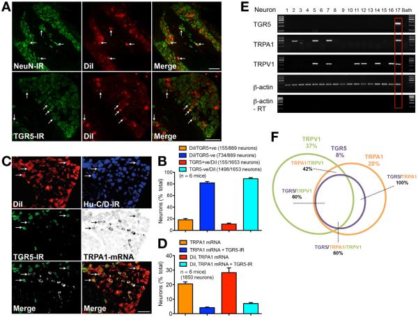
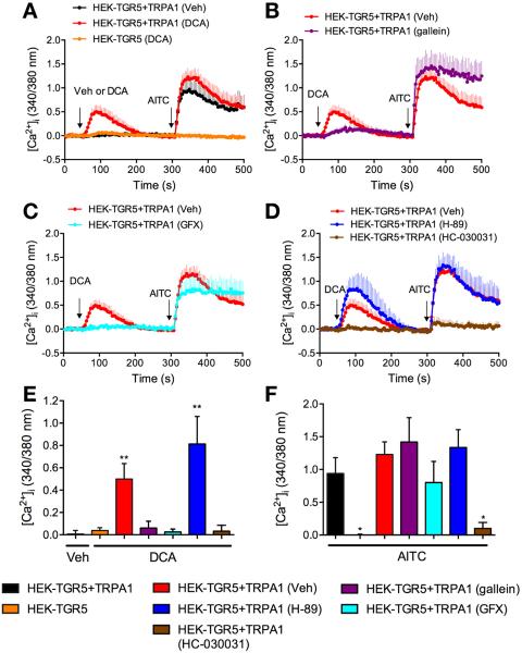
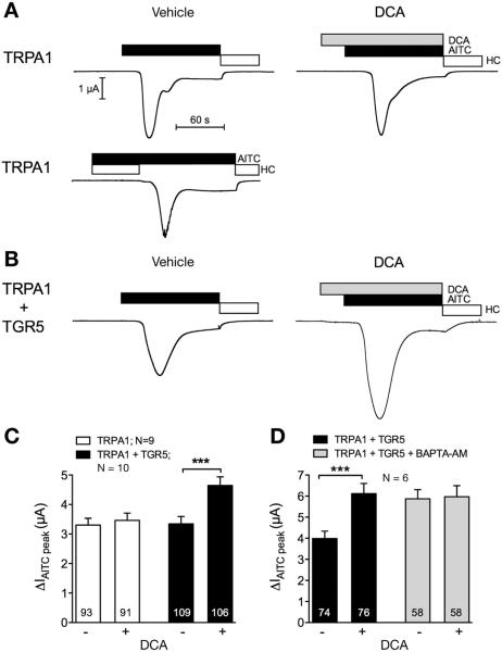
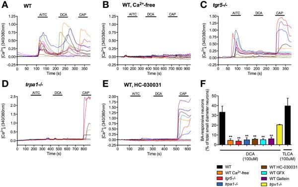
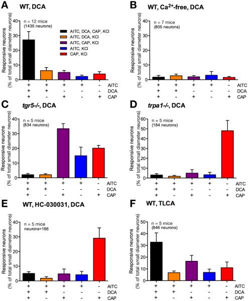
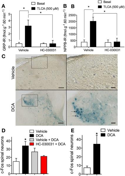
Methods: Co-expression of TGR5 and TRPA1 in cutaneous afferent neurons isolated from mice was analyzed by immunofluorescence, in situ hybridization, and single-cell polymerase chain reaction. TGR5-induced activation of TRPA1 was studied in in HEK293 cells, Xenopus laevis oocytes, and primary sensory neurons by measuring Ca(2+) signals. The contribution of TRPA1 to TGR5-induced release of pruritogenic neuropeptides, activation of spinal neurons, and scratching behavior were studied using TRPA1 antagonists or Trpa1(-/-) mice.

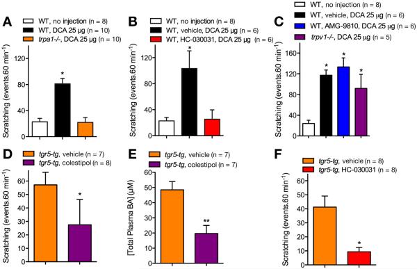
Results: TGR5 and TRPA1 protein and messenger RNA were expressed by cutaneous afferent neurons. In HEK cells, oocytes, and neurons co-expressing TGR5 and TRPA1, BAs caused TGR5-dependent activation and sensitization of TRPA1 by mechanisms that required Gβγ, protein kinase C, and Ca(2+). Antagonists or deletion of TRPA1 prevented BA-stimulated release of the pruritogenic neuropeptides gastrin-releasing peptide and atrial natriuretic peptide B in the spinal cord. Disruption of Trpa1 in mice blocked BA-induced expression of Fos in spinal neurons and prevented BA-stimulated scratching. Spontaneous scratching was exacerbated in transgenic mice that overexpressed TRG5. Administration of a TRPA1 antagonist or the BA sequestrant colestipol, which lowered circulating levels of BAs, prevented exacerbated spontaneous scratching in TGR5 overexpressing mice.

Conclusions: BAs induce pruritus in mice by co-activation of TGR5 and TRPA1. Antagonists of TGR5 and TRPA1, or inhibitors of the signaling mechanism by which TGR5 activates TRPA1, might be developed for treatment of cholestatic pruritus.

Keywords: Itching; Liver; Mouse Model; Signal Transduction.


Figures

Figure 1
Figure 1
Localization and expression of TGR5-IR, NeuN-IR, and TRPA1 mRNA in cutaneous afferent neurons. (A) Co-localization of NeuN-IR and TGR5-IR in DiI-positive cutaneous afferent DRG neurons (arrows). Scale bar = 100 μm. (B) Proportion of DiI-positive neurons with (TGR5-positive) or without (TGR5-negative) co-expression of TGR5-IR (889 or 1653 neurons, 6 mice). (C) Co-localization of Hu-C/D-IR, TGR5-IR, and TRPA1 mRNA in DiI-positive cutaneous afferent DRG neurons (arrows). Merged images show TGR5-IR + TRPA1 mRNA (left) and DiI + TGR5-IR + TRPA1 mRNA (right). Scale bar = 100 μm. (D) Proportion all neurons or of DiI-positive neurons expressing TGR5-IR alone or TGR5-IR + TRPA1 mRNA (1850 neurons, 6 mice). (E) Single-cell reverse transcription polymerase chain reaction of small-diameter, DiI-positive, cutaneous afferent neurons. Transcripts of TGR5, TRPA1, TRPV1, and β-actin were amplified. Results from 17 neurons are shown (60 neurons, 6 mice). Red box denotes co-expression of TGR5, TRPA1, and TRPV1. RT, reverse transcriptase. (F) Venn diagram indicating the proportion of small-diameter, Di-positive cutaneous afferent neurons expressing TRG5, TRPA1, and TRPV1.
Figure 2
Figure 2
BA and TGR5-induced activation and sensitization of TRPA1 in HEK293 cells. (AD). [Ca2+]i measured in HEK-TGR5 and HEK-TGR5+TRPA1 cells treated with vehicle (Veh) or DCA (100 μM) and then AITC (100 μM). Some cells were treated with gallein (B), GF-109203X (GFX, C), H-89 or HC-030031 (D). (E, F) Pooled results showing maximal increase in [Ca2+]i over basal. Triplicate measurements, 3–8 independent experiments; *P < .05 compared with vehicle (E) or AITC (F) in HEK-TGR5 + TRPA1 cells. **P < .001.
Figure 3
Figure 3
BA and TGR5-induced activation of TRPA1 currents in Xenopus laevis oocytes. Oocytes expressing TRPA1 alone or TRPA1 and TGR5 were stimulated with the TRPA1 agonist AITC (50 μM, black bar) and the TRPA1 antagonist HC-030031 (open bar). TGR5 was activated by DCA (500 μM, gray bar). (A, B) Representative whole-cell current traces of oocytes expressing TRPA1 (A) or TRPA1 and TGR5 (B). Oocytes were treated with vehicle or DCA 30 seconds before AITC. (C, D) Change in AITC currents (ΔIAITC peak) in oocytes treated with DCA (+) or vehicle (−) (C). ΔIAITC peak in oocytes preincubated for 3 hours in BAPTA-AM or vehicle and subsequently treated with DCA (+) or vehicle (−) (D). Numbers inside the columns indicate the number of individual oocytes measured. N indicates the number of batches of oocytes. ***P < .001.
Figure 4
Figure 4
TGR5- and TRPA1-dependent BA signaling in DRG neurons. [Ca2+]i was measured in small diameter neurons from wild-type (WT) mice (A), WT mice in Ca2+-free medium (B), tgr5−/− mice (C), trpa1−/− mice (D), and WT mice treated with the TRPA1 antagonist HC-030031 (E). They were challenged sequentially with AITC (100 μM), DCA (100 μM) and capsaicin (CAP, 1 μM). Responses of KCl (50 mM)-responsive neurons are shown. (F) Pooled data showing the proportion of DCA- or TLCA-responsive neurons in the different experimental groups. In (AE), each line represents a single neuron. (F) Pooled data from 2281 neurons, n = 17 mice. **P < .01 to DCA in WT.
Figure 5
Figure 5
The proportion of DRG neurons responding to BAs and agonists of TRP channels. [Ca2+]i was measured in small diameter neurons from wild-type (WT) mice (A, F), WT mice in Ca2+-free medium (B), tgr5−/− mice (C), trpa1−/− mice (D), WT mice treated with the TRPA1 antagonist HC-030031 (E). They were challenged sequentially with AITC (100 μM), DCA (AE) or TLCA (F) (both 100 μM), capsaicin (CAP, 1 μM), and finally KCl (50 mM). The proportion of small-diameter neurons that responded to the combinations of AITC, DCA or TLCA, and capsaicin are shown. Pooled data from 184–1435 neurons, n = 5–12 mice.
Figure 6
Figure 6
Contributions of TRPA1 to BA-evoked release of pruritogenic neuropeptides and activation of spinal neurons. (A, B) Release of GRP-IR (A) and NPPB-IR (B) from superfused slices of rat spinal cord under basal conditions and after stimulation with TLCA (500 μM, 60 minutes). Tissues were preincubated with vehicle or HC-030031 (50 μM). n = 4; *P < .05. (CE) Expression of c-fos in neurons in the dorsal horn of c-fos reporter mice. (C) Photomicrographs of the dorsal horn of the spinal cord (C1–C7) 60 minutes after injection of DCA (25 μg, SC) or vehicle to the nape of the neck of conscious mice. Scale bar, left panel = 100 μm; right panel = 25 μm. (D) Quantification of c-fos–expressing neurons in superficial laminae of the spinal cord (C1–C7) 60 minutes after injection of DCA or vehicle. (E) Quantification of c-fos–expressing neurons 60 minutes after injection of DCA or vehicle in anesthetized mice. Subgroups of the DCA-treated mice were pretreated with HC-030031 or vehicle 30 minutes before DCA. n = 3 mice; *P < .05 to vehicle.
Figure 7
Figure 7
Contributions of TRPA1 to scratching evoked by exogenous and endogenous BAs. (AC) DCA-evoked scratching in wild-type (WT) and trpa1−/− mice. DCA was injected to the nape of the neck (25 μg SC) and scratching was measured for 60 minutes in WT mice and trpa1−/− mice (A), in wild-type mice pretreated with HC-030031 or vehicle (B), and in wild-type mice pretreated with AMG-9810 or vehicle and in trpv1−/− mice (C). (DF) Scratching and serum BAs levels in tgr5-tg mice. tgr5-tg mice were treated with colestipol or vehicle for 5 days before measurement of scratching (D) and plasma concentrations of total BAs (E). Scratching was also measured in tgr5-tg mice pretreated with HC-030031 or vehicle (F). n = 6–10 mice; *P < .05, **P < .01 to untreated wild-type mice (AC) or vehicle (DF).

References

    1. Ikoma A, Steinhoff M, Stander S, et al. The neurobiology of itch. Nat Rev Neurosci. 2006;7:535–547. - PubMed
    1. Imamachi N, Park GH, Lee H, et al. TRPV1-expressing primary afferents generate behavioral responses to pruritogens via multiple mechanisms. Proc Natl Acad Sci U S A. 2009;106:11330–11335. - PMC - PubMed
    1. Liu Q, Tang Z, Surdenikova L, et al. Sensory neuron-specific GPCR Mrgprs are itch receptors mediating chloroquine-induced pruritus. Cell. 2009;139:1353–1365. - PMC - PubMed
    1. Steinhoff M, Neisius U, Ikoma A, et al. Proteinase-activated receptor-2 mediates itch: a novel pathway for pruritus in human skin. J Neurosci. 2003;23:6176–6180. - PMC - PubMed
    1. Wilson SR, Gerhold KA, Bifolck-Fisher A, et al. TRPA1 is required for histamine-independent, Mas-related G protein-coupled receptor-mediated itch. Nat Neurosci. 2011;14:595–602. - PMC - PubMed

Publication types

MeSH terms