. 2014 Oct;16(10):1016-26, 1-8.
doi: 10.1038/ncb3028. Epub 2014 Sep 7.

Systematic characterization of deubiquitylating enzymes for roles in maintaining genome integrity

Affiliations

Systematic characterization of deubiquitylating enzymes for roles in maintaining genome integrity

Ryotaro Nishi et al. Nat Cell Biol. 2014 Oct.

Abstract

DNA double-strand breaks (DSBs) are perhaps the most toxic of all DNA lesions, with defects in the DNA-damage response to DSBs being associated with various human diseases. Although it is known that DSB repair pathways are tightly regulated by ubiquitylation, we do not yet have a comprehensive understanding of how deubiquitylating enzymes (DUBs) function in DSB responses. Here, by carrying out a multidimensional screening strategy for human DUBs, we identify several with hitherto unknown links to DSB repair, the G2/M DNA-damage checkpoint and genome-integrity maintenance. Phylogenetic analyses reveal functional clustering within certain DUB subgroups, suggesting evolutionally conserved functions and/or related modes of action. Furthermore, we establish that the DUB UCHL5 regulates DSB resection and repair by homologous recombination through protecting its interactor, NFRKB, from degradation. Collectively, our findings extend the list of DUBs promoting the maintenance of genome integrity, and highlight their potential as therapeutic targets for cancer.


Figures

Figure 1
Figure 1. Screen to identify DUBs connected to DSB repair or the DNA damage G2/M checkpoint
Schematic representation of the screen for human DUBs involved in DSB responses. In the primary screen, GFP-fused DUB constructs were transfected into cells stably expressing RFP-fused 53BP1, then localisation of GFP-DUBs to sites of DNA damage induced by laser micro-irradiation was examined. In parallel, each DUBs was depleted by siRNA pools and subjected to immunoblotting analysis for DDR or G2/M checkpoint markers. In the secondary screen, 44 DUBs obtained from the primary screen were subjected to neutral comet assay after depletion of each DUBs by siRNA pools.
Figure 2
Figure 2. Classification of screen results
(a) Results of screen for DUBs involved in G2/M DNA-damage checkpoint. Ratio of signal intensity of phosphorylated histone H3 Ser-10 (H3 pS10) normalized to total histone H3 level before and after damage (phleomycin 40 μg/ml, 2 h) is plotted. Each DUB is numbered in ascending order based on the H3 pS10 level, with the names of the corresponding DUBs provided in Supplementary Table 1. Data shown is the one experiment carried out for each DUB depletion and the mean of ten experiments for siRNA control. Numbers 74-90, which are coloured with green or red, are: CSN6, STAMBP, USP6, HINL1, USP8, EIF3S3, USP52, UCHL1, CSN5, USP20, USP49a, USP19, USPL1, PSMD14, USP29, UCHL3 and USP37. (b) Results of DSB repair secondary screen with indicated DUB siRNAs. Repair efficiencies were determined by the tail moment ratio between 2 h after phleomycin (40 μg/ml) removal (recovery) and immediately after treatment (damaged) Data show the means of two (DUB, XRCC4 and BRCA1 depletions) or seven (siRNA control depletions) biologically independent experiments for respectively. (a, b) Two and three times standard deviation of siRNA control (2 × and 3 × SD, respectively) are indicated. Depletion of XRCC4 and BRCA1 are supplied as positive controls. (c) Tail moments (arbitrary unit: AU) of cells transfected with indicated siRNAs without exogenous DNA-damage were plotted. Data show the means of two (DUB, XRCC4 and BRCA1 depletions) or seven (siRNA control) biologically independent experiments respectively. One times standard deviation of siRNA control is indicated by horizontal blue line.
Figure 3
Figure 3. Classification of DUBs based on localisation, DSB repair defects and phylogenetic analysis
(a) Classification of screen results based on localisation of GFP-DUBs to laser micro-irradiation sites and effects on DSB repair using siRNA pools (neutral comet assay). DUBs in bold represent DUBs with previously reported connections to the DDR. *: USP42 that was excluded from sites of DNA damage. **: not tested for localisation. (b, c) Phylogenetic analysis of human DUBs for (c) DSB repair or (d) G2/M checkpoint. DUBs are coloured green or red based on a degree of defect as shown in a and b. Human DUBs are classified into five subfamilies; ubiquitin-specific proteases (USPs), ubiquitin carboxyl-terminal hydrolases (UCHs), ovarian tumor proteases (OTUs), Machado-Joseph disease enzymes (MJDs) and JAB1/MPN/MOV34 metalloenzymes (JAMMs) (See main text introduction for details).
Figure 4
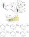
Figure 4. Verification of screen results
(a) Three-dimensional scatter plot of screen results. Localisation to DNA damage sites is divided into three categories on the x-axis (no effect, recruited or excluded). Immunoblotting screen results were scored based on the numbers of altered phosphorylation signals (0, 1, 2 and 3). For the DSB repair assay, tail moment ratio is plotted on the z-axis. DUBs are coloured based on comet assay DNA repair defects, as indicated by the bar on the left. (b) Neutral comet assays with two individual siRNAs targeting indicated DUBs. Data show the means of two (DUB depletions) or three (control siRNA) biologically independent experiments respectively. The blue line indicates two times standard deviation of siRNA control. DUBs that scored positive in all three screens are shown in blue. (c) Clonogenic survival assays with individual siRNAs targeting top hit DUBs from the screen. Data represent the individual results of two biologically independent experiments (solid lines and dashed lines).
Figure 5
Figure 5. UCHL5 promotes HR repair
(a) Left: Clonogenic survival assays with IR. Depletions of XRCC4 (siXRCC4) and BRCA1 (siBRCA1) are positive controls. Data show the means of four biologically independent experiments. The error bars indicate standard error of means. Right: Depletion efficiency of UCHL5 with siRNAs targeting the coding sequence (#1 and #2) or 3′UTR. Tubulin is shown as a loading control. (b, c) U2OS cells or U2OS cells carrying a direct repeat-GFP reporter transfected with the indicated siRNAs were processed for NHEJ (b) or HR (c) repair assays; Ligase IV depletion (siLigIV) and CtIP depletion (siCtIP) are respective positive controls. Data show the means of two (b) or three (c) biologically independent experiments, respectively. p-values are indicated by asterisks (**; p < 0.005, ***; p < 0.001). The error bars indicate standard error of means. (d) GFP or GFP-UCHL5 (wild-type: WT or deubiquitylase dead: DD) expressing U2OS cells transfected with the indicated siRNAs were processed for neutral comet assays. Data represent the means of two biologically independent experiments. (e) U2OS cells stably expressing RFP-53BP1 were transiently transfected with GFP-UCHL5 (WT or DD) and subjected to laser micro-irradiation. Images were taken before (undamaged: UD) or 15 min after irradiation (damaged: D). Localisation of endogenous UCHL5 to site of DNA damage was not examined due to lack of a suitable antibody. Arrows indicate irradiated areas. Scale bar indicates 10 μm.
Figure 6
Figure 6. UCHL5 contributes to resection by regulating EXO1 recruitment
(a, b, c) Cells transfected with indicated siRNAs were treated with camptothecin (CPT, 1 μM, 1 h) then subjected to immunoblotting with indicated antibodies (a), quantitative resection assay with anti-BrdU antibody (the means of three biologically independent experiments with standard errors of means, * means p < 0.05) (b), or anti-RPA antibody (data represent the means of two biologically independent experiments) (c). (d, e) GFP or GFP-UCHL5 (WT or DD) expressing U2OS cells transfected with indicated siRNAs were treated or mock treated with 1 μM of CPT for 1 h and analyzed by immunoblotting as indicated. (f) Intensity of GFP-CtIP at DNA damage sites relative to the unirradiated area was quantified 15 min after irradiation. Data show the means of three biologically independent experiments with error bars indicating standard errors of means. (g) Kinetics of GFP-EXO1 accumulation at DNA damage sites was assessed in cells transfected with indicated siRNAs. Signal intensity of GFP-EXO1 at DNA damage sites relative to the unirradiated area was quantified. Data show the means of three biologically independent experiments with error bars indicating standard errors of means.
Figure 7
Figure 7. UCHL5 regulates resection by protecting NFRKB from proteasomal degradation
(a) Cells transfected with indicated siRNAs were processed for immunoblotting with indicated antibodies. Arrow indicates position of NFRKB. (b) Left: Cells transfected with indicated siRNAs were incubated with 100 μg/ml of cycloheximide (CHX) for various times and processed for immunoblotting with indicated antibodies. Right: Quantification of data shown on left. Data show the means of two biologically independent experiments. (c) GFP or GFP-UCHL5 expressing cells transfected with indicated siRNAs were analyzed by immunoblotting as indicated. (d) GFP and GFP-NFRKB expressing cells were mock transfected or transiently transfected with an expression plasmid of HA-tagged ubiquitin (HA-Ub). Chromatin fractions were immunoprecipited with an anti-GFP antibody followed by immunoblotting. Brackets indicate ubiquitylated NFRKB. For inputs, see Supplementary Fig. 3f. (e) GFP or GFP-NFRKB stably expressing cells transfected with indicated siRNAs were subjected to quantitative resection assays. Data show the means of two biologically independent experiments. (f) Modified “traffic light reporter system” based HR assay with indicated siRNAs. Data show the means of two biologically independent experiments. (g) U2OS cells transfected with indicated siRNAs were subjected to clonogenic survival assays after IR. Data show the means of two biologically independent experiments. (h) Cells transfected with individual or indicated combinations of siRNAs were processed for quantitative resection assays. Data show the means of two biologically independent experiments. (i) U2OS cells transfected with indicated siRNAs were subjected to quantitative resection assays. Data show the means of two biologically independent experiments. (j) U2OS cells were incubated with CHX (100 μg/ml) and or MG132 (10 μM) for 1 h prior to camptothecin (CPT) treatment (1 μM, 1 h). Nucleoplasmic fractions were subjected to immunoblotting with indicated antibodies. Relative protein levels of NFRKB are indicated with normalization by HDAC1 levels. (k) After incubating GFP or GFP-NFRKB expressing cells with 10 μM MG132, nucloplasmic (Nu) and chromatin (Ch) fraction were immunoprecipitated with anti-GFP antibody followed by immunoblotting. For inputs, see Supplementary Fig. 4l.

References

    1. Hoeijmakers JH. Genome maintenance mechanisms for preventing cancer. Nature. 2001;411:366–374. - PubMed
    1. Khanna KK, Jackson SP. DNA double-strand breaks: signaling, repair and the cancer connection. Nat Genet. 2001;27:247–254. - PubMed
    1. Kastan MB. DNA damage responses: mechanisms and roles in human disease: 2007 G.H.A. Clowes Memorial Award Lecture. Mol Cancer Res. 2008;6:517–524. - PubMed
    1. Shiloh Y. The ATM-mediated DNA-damage response: taking shape. Trends Biochem Sci. 2006;31:402–410. - PubMed
    1. Jackson SP. Sensing and repairing DNA double-strand breaks. Carcinogenesis. 2002;23:687–696. - PubMed

Publication types

MeSH terms