. 2014 Sep;70(Pt 9):2301-15.
doi: 10.1107/S1399004714013777. Epub 2014 Aug 29.

Latent and active abPPO4 mushroom tyrosinase cocrystallized with hexatungstotellurate(VI) in a single crystal

Affiliations

Latent and active abPPO4 mushroom tyrosinase cocrystallized with hexatungstotellurate(VI) in a single crystal

Stephan Gerhard Mauracher et al. Acta Crystallogr D Biol Crystallogr. 2014 Sep.

Abstract

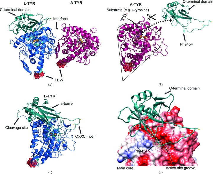
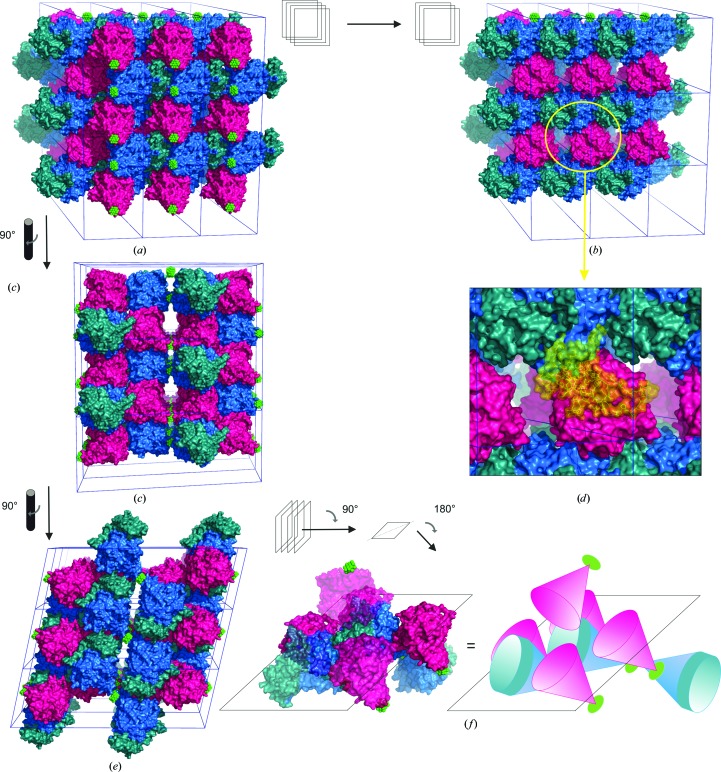
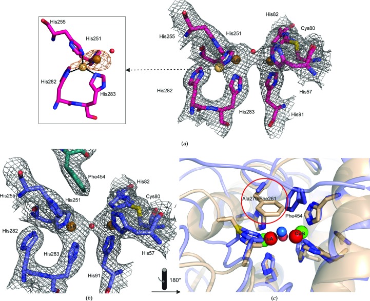
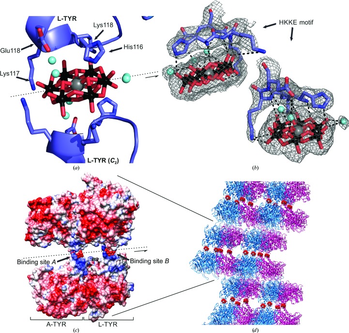
Tyrosinases, bifunctional metalloenzymes, catalyze the oxidation of monophenols and o-diphenols to o-quinones, the precursor compounds of the brown-coloured pigment melanin. In eukaryotic organisms, tyrosinases are expressed as latent zymogens that have to be proteolytically cleaved in order to form highly active enzymes. This activation mechanism, known as the tyrosinase maturation process, has scientific and industrial significance with respect to biochemical and technical applications of the enzyme. Here, not only the first crystal structure of the mushroom tyrosinase abPPO4 is presented in its active form (Ser2-Ser383) and in its 21 kDa heavier latent form (Ser2-Thr545), but furthermore the simultaneous presence of both forms within one single-crystal structure is shown. This allows for a simple approach to investigate the transition between these two forms. Isoform abPPO4 was isolated and extensively purified from the natural source (Agaricus bisporus), which contains a total of six polyphenol oxidases (PPOs). The enzyme formed crystals (diffracting to a resolution of 2.76 Å) owing to the employment of the 6-tungstotellurate(VI) salt (Na6[TeW6O24]·22H2O) as a cocrystallization agent. Two of these disc-shaped Anderson-type polyoxoanions [TeW6O24](6-) separate two asymmetric units comprising one crystallographic heterodimer of abPPO4, thus resulting in very interesting crystal packing.

Keywords: Agaricus bisporus; abPPO4; polyphenol oxidase; tyrosinase.


Figures

Figure 1
Figure 1
Overall structure. Colour code: L-TYR main core, blue; C-terminal domain, turquoise; A-TYR, purple; sodium ions, yellow; copper ions, bronze; POM (TEW) oxygen, red; tellurium, grey; positive electrostatic potential of coulombic surface, blue; negative electrostatic potential of coulombic surface, red. (a) Overall structure of asymmetric unit; the crystallographic heterodimer of abPPO4. L-TYR is shown on the left and A-TYR is depicted on the right. (b) Illustration of the activation process. By the proteolytic removal of the C-terminal domain the active site becomes solvent-exposed and substrates (e.g. l-tyrosine) are able to approach. (c) Different viewing angle of the single monomer of L-TYR to illustrate the β-barrel-shaped C-terminal domain, the proteolytic cleavage site and the putative CXXC motif-containing copper grappler. (d) Coulombic surface illustration of the L-TYR main core with the C-terminal domain (cartoon) attached. The solvent-accessible groove, exhibiting a strong negative electrostatic potential, is indicated by the dashed arrow. Phe454 is shown to protrude into the active site (transparent surface around the active site).
Figure 2
Figure 2
Multiple sequence alignment of abPPO4 (UniProt K9I869), aoTYR (UniProt B8NJ95) and abPPO3 (UniProt C7FF04). Colour code for secondary-structure elements: α-helices, blue tubes; β-strands, green arrows; loops, red arrows; hydrogen-bonded turnovers, TT. Colour code for highlighted residues: copper-binding histidines, orange; conserved tyrosinase motifs, yellow; mutations discerning abPPO4 K9I869 from abPPO4 C7FF04, purple; putative transmembrane anchor (Ala569–Ala591), raspberry; acetylated N-terminus, green.
Figure 3
Figure 3
Crystal packing. Colour code: L-TYR main core, blue; C-terminal domain, turquoise; A-TYR, purple; POM (TEW), green. (a) Crystal packing in 1 × 3 × 3 supercell. Four layers of protein (surface illustration) pointing alternately in opposing directions. The front layer exposes its POM side (front). (b) The same view as in (a) but with the first layer removed. Hence, the protein side is exposed (back). Here, the head-to-body alignment of L-TYR is discernible. (c) The image in (a) rotated by 90°, showing the sandwich-like layer stacking. (d) The insert shows a C-terminal domain attached notionally to A-TYR and clearly clashing with L-TYR of the adjacent asymmetric unit. (e) The image in (c) rotated by 90°. (f) The image in (e) tilted by 90° and rotated by 180°. One central L-TYR chain surrounded by the vicinal flanking chains (5× A-TYR, 2× L-TYR). An equivalent schematic illustration is shown on the right.
Figure 4
Figure 4
Superimpositions. Colour code: L-TYR main core, blue; L-TYR C-terminal domain, turquoise; A-TYR main core, purple; aoTYR main core, green; aoTYR C-terminal domain, lime green; abPPO3 main core, orange; abPPO3 light subunit, yellow; L-TYR abPPO4 copper ions, green; respective superimposed copper ions, red; sodium ions, yellow; water molecules, blue; tropolone, pink; POM oxygen spheres, red. (a) Superimposition of L-TYR abPPO4 with A-TYR abPPO4. (b) Magnified region of loopX (Asn236–Ser246) which is pushed sideways by the attachment of the C-terminal domain. The dashed arrow indicates the motion of loopX owing to the removal of the C-terminal domain. A sodium ion occupying the respective metal-binding site of loopX in L-TYR is thereby lost. (c) Magnified superimposition of the respective active sites (A-TYR and L-TYR). (d) Superimposition of L-TYR abPPO4 with A-TYR abPPO3. (e) Magnified superimposition of the respective active sites (L-TYR abPPO4 and A-TYR abPPO3) depicting a similar location of the placeholder Phe454 and the inhibitor tropolone. (f) Superimposed POM-binding region of L-TYR abPPO4 and abPPO3 showing that no conformational change is induced owing to POM binding. (g) Superimposition of L-TYR abPPO4 with aoTYR. The two loops putatively pushed sideways owing to the attachment of the C-terminal domain of aoTYR are indicated as loopX and loopY, respectively. The insert shows the superimposed proteolytic cleavage site located on the back side in the respective perspective of the main view. In contrast to an α-helical location in aoTYR, the site is located on a loop in abPPO4. (h) Parallel arranged C-terminal subunits of L-TYR abPPO4 (left) and aoTYR (right) for a proper comparison. The dashed line indicates missing residues. (i) Magnified superimposition of the respective active sites (L-TYR abPPO4 and aoTYR).
Figure 5
Figure 5
Active site of L-TYR and A-TYR abPPO4. Colour code: A-TYR, purple; L-TYR, blue; copper ion, bronze (alternative conformation, light bronze); 2mF omF c electron-density mesh (0.63 e Å−3, 1.5 r.m.s.d.), grey; anomalous electron-density mesh (0.40 e Å−3, 3 r.m.s.d.), orange; catechol oxidase from I. batatas, beige; L-TYR abPPO4 copper ions, green; respective superimposed ibCO copper ions, red. (a) Active site of A-TYR lacking the protruding Phe454 (C-terminal domain) with a copper distance of 4.2 Å. A water molecule (Wat128, ocupancy 0.90, B = 42.65 Å2) is bridging the two copper ions in a triangular position (CuA–Wat = 2.4 Å; Wat–CuB = 2.3 Å). CuB shows an alternative conformation in the vicinity of His282 (CuB1–CuB2 = 2.3 Å). His255, His251, His282 and His283 are coordinating the weakly occupied alternative CuB site (CuB1, occupancy 0.92, B = 30.62 Å2; CuB2, occupancy 0.08, B = 29.31 Å2) in a tetrahedral conformation (Cu—N∊/δ = 2.1–2.2 Å). The inset shows the anomalous scattering electron-density map, which clearly exhibits a hump towards His282. Thus, positional flexibility of the CuB site is indicated. Notably, His282 in (a) is side-chain flipped compared with (b). (b) Latent active site of L-TYR showing defined electron density at both copper-binding sites. A water molecule (Wat129, occupancy 0.95, B = 32.90 Å2) is bridging the copper (CuA–CuB = 4.7 Å) spheres positioned directly on a line between the two ions (CuA–Wat = 2.3 Å; Wat–CuB = 2.3 Å). (c) Superimposition of the active site of L-TYR abPPO4 and catechol oxidase from I. batatas. The CuA site-blocking bulky residue (Phe261) in ibCO is substituted by the less bulky alanine residue (Ala270) in L-TYR abPPO4.
Figure 6
Figure 6
POM (TEW) binding site. Colour code: L-TYR, blue; A-TYR, purple; O atoms, red; Te atom, grey, W atoms, black; water molecules, cyan; hydrogen bonds, dashed black lines; positively charged coulombic surface, blue; negatively charged coulombic surface, red. (a) POM-binding (L-TYR) site B is located at the tip (start of α-helix α5) of the conical-shaped main core. The polyoxoanion lies on a twofold axis (C 2) embedded by two opposing HKKE motifs (L-TYR and L-TYR C 2). (b) Image of two different views of the one-sided POM-binding site B with the respective electron density indicated by a grey mesh (2mF omF c, 0.40 e Å−3, 1 r.m.s.d.) and all possible hydrogen bonds outlined. (c) Coulombic surface image of two opposing asymmetric units (heterodimer, A-TYR/L-TYR abPPO4) sharing two polyoxoanions on a twofold axis. This image illustrates how the POM acts as a conjunction linking two otherwise repulsive surfaces. (c) Image section of crystal packing displaying a sandwich-like constitution (protein layer–POM layer–protein layer).

References

    1. Adams, P. D. et al. (2010). Acta Cryst. D66, 213–221.
    1. Bourquelot, E. & Bertrand, G. (1895). C. R. Soc. Biol. 47, 582–584.
    1. Chen, V. B., Arendall, W. B., Headd, J. J., Keedy, D. A., Immormino, R. M., Kapral, G. J., Murray, L. W., Richardson, J. S. & Richardson, D. C. (2010). Acta Cryst. D66, 12–21. - PMC - PubMed
    1. Corey, R. B., Stanford, R. H., Marsh, R. E., Leung, Y. C. & Kay, L. M. (1962). Acta Cryst. 15, 1157–1163.
    1. Cuff, M. E., Miller, K. I., van Holde, K. E. & Hendrickson, W. A. (1998). J. Mol. Biol. 278, 855–870. - PubMed

Publication types

Associated data