. 2014 Sep;70(Pt 9):2444-54.
doi: 10.1107/S1399004714015053. Epub 2014 Aug 29.

Structure of the 2,4'-dihydroxyacetophenone dioxygenase from Alcaligenes sp. 4HAP

Affiliations

Structure of the 2,4'-dihydroxyacetophenone dioxygenase from Alcaligenes sp. 4HAP

R Keegan et al. Acta Crystallogr D Biol Crystallogr. 2014 Sep.

Abstract

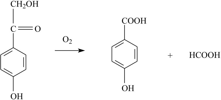
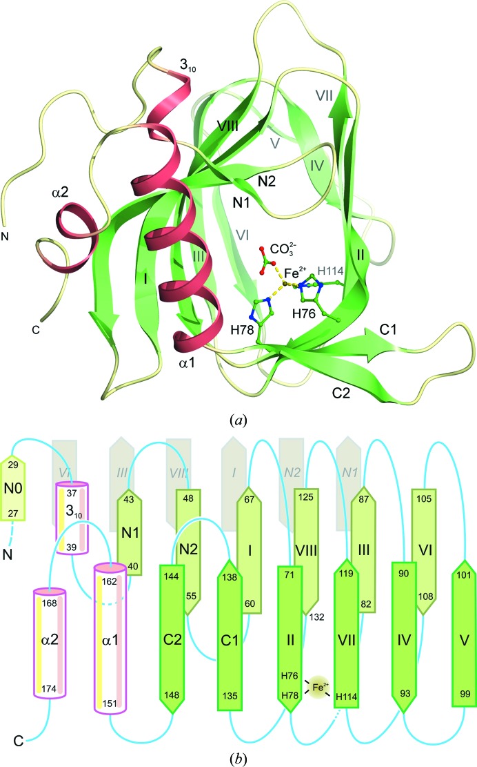
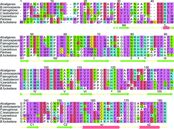
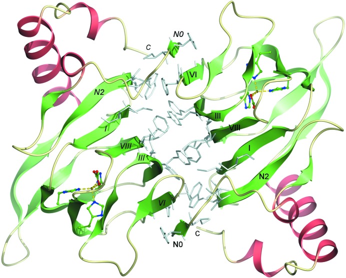
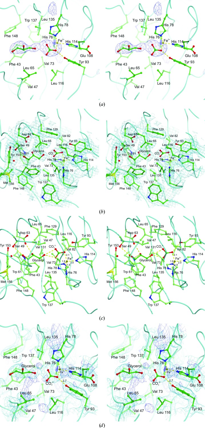
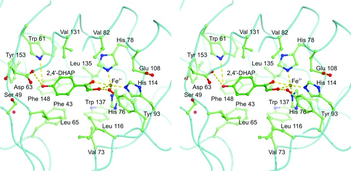
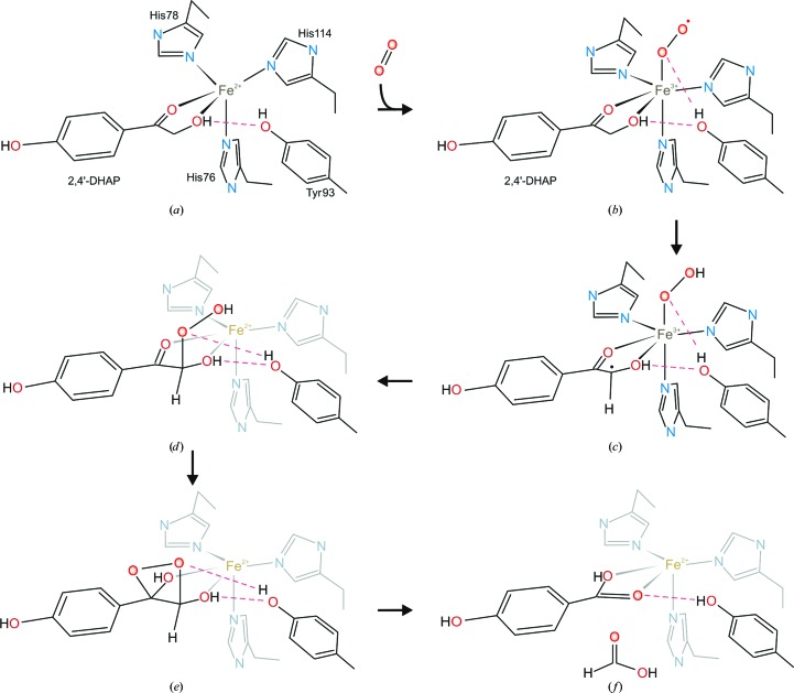
The enzyme 2,4'-dihydroxyacetophenone dioxygenase (DAD) catalyses the conversion of 2,4'-dihydroxyacetophenone to 4-hydroxybenzoic acid and formic acid with the incorporation of molecular oxygen. Whilst the vast majority of dioxygenases cleave within the aromatic ring of the substrate, DAD is very unusual in that it is involved in C-C bond cleavage in a substituent of the aromatic ring. There is evidence that the enzyme is a homotetramer of 20.3 kDa subunits, each containing nonhaem iron, and its sequence suggests that it belongs to the cupin family of dioxygenases. In this paper, the first X-ray structure of a DAD enzyme from the Gram-negative bacterium Alcaligenes sp. 4HAP is reported, at a resolution of 2.2 Å. The structure establishes that the enzyme adopts a cupin fold, forming dimers with a pronounced hydrophobic interface between the monomers. The catalytic iron is coordinated by three histidine residues (76, 78 and 114) within a buried active-site cavity. The iron also appears to be tightly coordinated by an additional ligand which was putatively assigned as a carbonate dianion since this fits the electron density optimally, although it might also be the product formate. The modelled carbonate is located in a position which is highly likely to be occupied by the α-hydroxyketone group of the bound substrate during catalysis. Modelling of a substrate molecule in this position indicates that it will interact with many conserved amino acids in the predominantly hydrophobic active-site pocket where it undergoes peroxide radical-mediated heterolysis.

Keywords: catalytic mechanism; cupin fold; dioxygenase; iron binding.


Figures

Figure 1
Figure 1
The reaction catalysed by 2,4′-dihydroxyacetophenone dioxygenase (DAD). The enzyme has a high affinity for oxygen, which is used for C—C bond cleavage of the substrate (2,4′-dihydroxyacetophenone), yielding 4-hydroxybenzoic acid and formate.
Figure 2
Figure 2
The tertiary structure of Alcaligenes DAD. (a) The fold of the monomer viewed along the cupin barrel of the enzyme towards the iron-binding site which is formed by three invariant histidine residues (76, 78 and 114) and a putative carbonate ligand (shown in ball-and-stick representation with the iron coloured rust brown and its dative bonds as yellow dashes). Regions of β-strand are coloured pale green and the helical segments are shown in clay-red with the intervening loops in beige. Note that strand N0 partakes in one of the cupin-barrel sheets of the neighbouring monomer in the dimer. (b) A topology diagram of DAD showing the cupin-barrel sheets with residue numbers for all of the secondary-structure elements. The iron-binding site formed by His76, His78 and His114 in strands II and VII is shown, as is the involvement of strand N0 in the abutting β-sheet of the neighbouring monomer (pale grey stands at the back, labelled in italics).
Figure 3
Figure 3
A sequence alignment of putative DAD enzymes. The secondary structure of the Alcaligenes sp. 4HAP enzyme is shown below the sequences. The amino-acid residues are colour-coded as follows: cyan, basic; red, acidic; green, neutral polar; pink, bulky hydrophobic; white, Gly, Ala and Pro; yellow, Cys. The iron-binding histidine residues are indicated with asterisks, which also conveniently mark the two cupin motifs (Dunwell et al., 2004 ▶). Numbering refers to the combined alignment.
Figure 4
Figure 4
The structure of the DAD dimer. The dimer is viewed along the crystallographic twofold axis which relates the two monomers. The hydrophobic amino acids which form the central dimer interface are shown in pale grey. The secondary-structure elements forming this central β-barrel of the dimer, in which strand N0 crosses over from one subunit to the next, are labelled in roman and italic text to aid in distinguishing the two constituent monomers.
Figure 5
Figure 5
The substrate-binding pocket of DAD. (a) A stereoview of the omit map for the active-site region of the protein showing the positive difference electron density for the unknown ligand(s) contoured at 3.0 r.m.s. Our interpretation of this density as an iron-bound carbonate and a cryoprotectant molecule, glycerol, is shown along with the iron-binding histidines and other residues in the vicinity. A more complete view of the active-site residues and putative ligands with their refined electron density, contoured at 1.0 r.m.s., is shown in (b). Both ligands fit the electron density satisfactorily, make reasonable hydrogen bonds and refine reasonably well. The dative bonds to the iron ion and the hydrogen bonds made by the putative glycerol and carbonate moieties (dark green) are shown as yellow dashed lines in (c). The glycerol makes hydrogen bonds to the putative carbonate and to Ser49, Trp61 and Asp63, which are conserved residues. (d) shows more details of the iron ligands along with the electron density for the putative carbonate and glycerol. The refined 2F oF c map is shown in pale blue contoured at the same level as in (b) and the residual F oF c positive difference density, contoured at 2.5 r.m.s., is shown in dark blue. There is a feature of difference density connected with the iron and pointing in the direction of Glu108 which may represent the binding site of diatomic oxygen. Where shown, dative-bond or hydrogen-bond lengths are in Å.
Figure 6
Figure 6
X-band EPR spectra of DAD. (a) 4000 G field sweep (an asterisk indicates trace Cu2+ contamination). (b) A sweep showing the region from 500 to 2100 G, with g values for major lines indicated. (c) DAD with a fivefold molar excess of substrate 2,4′-DHAP added, with g values for major lines indicated; vertical and horizontal scales are as in (b). Experimental parameters: microwave power 0.5 mW, modulation amplitude 5 G, modulation frequency 100 kHz, temperature 10 K; (b) and (c) are the result of four co-added scans.
Figure 7
Figure 7
A stereoview of a model of the aromatic substrate 2,4′-DHAP in the active site. The 2,4′-DHAP (shown in dark green) occupies the positions of the putative glycerol and carbonate. The phenolic –OH group interacts with Asp63 and Trp61 by hydrogen bonds and the α-­hydroxyketone group of 2,4′-DHAP interacts with the active-site iron by dative bonding and with the side chain of Tyr93 by a hydrogen bond. Note that the side chain of Phe129 which, like Phe43, interacts with the substrate by ring stacking has been omitted from the foreground for clarity.
Figure 8
Figure 8
The catalytic mechanism of DAD. (a) shows the substrate complex, as in Fig. 7 ▶, and the following parts (bf) indicate the steps of the reaction proposed by Paria et al. (2012 ▶) which is consistent with our experimentally determined structure. For clarity, steps (df) show the active-site histidines and the iron greyed-out since they are in the background in this view. The scheme emphasizes the key role played by the –OH group of Tyr93 in the stabilizing the intermediates.

References

    1. Aik, W., McDonough, M. A., Thalhammer, A., Chowdhury, R. & Schofield, C. J. (2012). Curr. Opin. Struct. Biol. 22, 691–700. - PubMed
    1. Baker, N. A., Sept, D., Joseph, S., Holst, M. J. & McCammon, J. A. (2001). Proc. Natl Acad. Sci. USA, 98, 10037–10041. - PMC - PubMed
    1. Beaven, G., Bowyer, A., Erskine, P., Wood, S. P., McCoy, A., Coates, L., Keegan, R., Lebedev, A., Hopper, D. J., Kaderbhai, M. A. & Cooper, J. B. (2014). Acta Cryst. F70, 823–826. - PMC - PubMed
    1. Bugg, T. D., Ahmad, M., Hardiman, E. M. & Singh, R. (2011). Curr. Opin. Biotechnol. 22, 394–400. - PubMed
    1. Chavez, F. A., Banerjee, A. & Sljivic, B. (2011). On Biomimetics, edited by L. Pramatarova, ch. 1. Rijeka InTech. 10.5772/18262.

Publication types

Substances

Associated data