. 2014 Sep;70(Pt 9):1180-5.
doi: 10.1107/S2053230X14017026. Epub 2014 Aug 29.

X-ray structures of Nfs2, the plastidial cysteine desulfurase from Arabidopsis thaliana

Affiliations

X-ray structures of Nfs2, the plastidial cysteine desulfurase from Arabidopsis thaliana

Thomas Roret et al. Acta Crystallogr F Struct Biol Commun. 2014 Sep.

Abstract

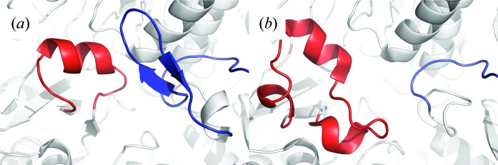
The chloroplastic Arabidopsis thaliana Nfs2 (AtNfs2) is a group II pyridoxal 5'-phosphate-dependent cysteine desulfurase that is involved in the initial steps of iron-sulfur cluster biogenesis. The group II cysteine desulfurases require the presence of sulfurtransferases such as SufE proteins for optimal activity. Compared with group I cysteine desulfurases, proteins of this group contains a smaller extended lobe harbouring the catalytic cysteine and have a β-hairpin constraining the active site. Here, two crystal structures of AtNfs2 are reported: a wild-type form with the catalytic cysteine in a persulfide-intermediate state and a C384S variant mimicking the resting state of the enzyme. In both structures the well conserved Lys241 covalently binds pyridoxal 5'-phosphate, forming an internal aldimine. Based on available homologous bacterial complexes, a model of a complex between AtNfs2 and the SufE domain of its biological partner AtSufE1 is proposed, revealing the nature of the binding sites.

Keywords: Arabidopsis thaliana; SUF machinery; cysteine desulfurase; iron–sulfur cluster.


Figures

Figure 1
Figure 1
Structure of AtNfs2. (a) Ribbon representation of AtNfs2. Protomers A and B are coloured green and blue, respectively. (b) Topology diagram of secondary structures. Helices (letters) are shown in red and β-strands (numbers) are shown in blue. The N-terminal and C-terminal extremities are shown in orange. (c) AtNfs2 monomer. The seven-stranded β-sheet, the four-stranded β-sheet and the β-hairpin are shown in yellow, red and purple, respectively. The extended lobe between β12 and β13 is in cyan. The PLP cofactor covalently bound to the Lys241 and the catalytic site Cys384 are shown as sticks.
Figure 2
Figure 2
Comparison between group I and group II cysteine desulfurases. (a) Active site of AtNfs2, a SufS-like group II CD. (b) Active site of IscS from A. fulgidus (PDB entry 4eb7), a group I CD. Monomers A and B are coloured white. The loop corresponding to a β-hairpin (in group II CDs) is coloured blue and the region containing the conserved cysteine residue (referred to as the ‘extended loop’) is coloured red.
Figure 3
Figure 3
Active site of AtNfs2. (a) Electron density around the catalytic residue (Cys384) in wild-type AtNfs2. The map shown is a σA-weighted 2mF oDF c map contoured at 1.2σ (0.44 e Å−3). (b) Electron density around Ser384 in the C384S variant. The map shown is a σA-weighted 2mF oDF c map contoured at 1.2σ (0.42 e Å−3). (c) Representation of the PLP-binding site with 2mF oDF c electron density (1.2σ and 0.44 e Å−3) around the cofactor and side chain of Lys241A. The PLP cofactor is covalently bound to Lys241A. Numerous residues are involved in the stabilization of the PLP cofactor, i.e. Thr110A, Ser238A, His240A and Thr292B for the phosphate group and Asp215A and Gln218A for the pyridine ring.
Figure 4
Figure 4
Cysteine desulfurase and sulfur-acceptor complexes. The CD–sulfurtransferase (SufE, CsdE and TusA) complexes and the CD–scaffold protein (IscU) complex are represented as ribbons. CDs are coloured red, yellow and green, and CD partners are coloured cyan, purple and pink. (a) Model of the Nfs2–SufEAtSufE1 complex from A. thaliana and the X-ray crystal structures of (b) CsdA–CsdE from E. coli, (c) Fe2S2 coordinated by three sulfurtransferase residues (Cys33, Cys58 and Cys102) and by one cysteine desulfurase residue (Cys321) in the IscS–IscU complex from A. fulgidus, (d) IscS–IscU from E. coli and (e) IscS–TusA from E. coli (PDB entries 4lw4, 4eb5, 3lvl and 3lvj, respectively).

References

    1. Adams, P. D. et al. (2010). Acta Cryst. D66, 213–221. - PubMed
    1. Chen, V. B., Arendall, W. B., Headd, J. J., Keedy, D. A., Immormino, R. M., Kapral, G. J., Murray, L. W., Richardson, J. S. & Richardson, D. C. (2010). Acta Cryst. D66, 12–21. - PMC - PubMed
    1. Collins, R., Johansson, A. L., Karlberg, T., Markova, N., van den Berg, S., Olesen, K., Hammarström, M., Flores, A., Schüler, H., Schiavone, L. H., Brzezinski, P., Arnér, E. S. & Högbom, M. (2012). PLoS One, 7, e30581. - PMC - PubMed
    1. Comeau, S. R., Gatchell, D. W., Vajda, S. & Camacho, C. J. (2004a). Bioinformatics, 20, 45–50. - PubMed
    1. Comeau, S. R., Gatchell, D. W., Vajda, S. & Camacho, C. J. (2004b). Nucleic Acids Res. 32, 96–99. - PMC - PubMed