. 2014 Sep;70(Pt 9):1186-92.
doi: 10.1107/S2053230X14017087. Epub 2014 Aug 29.

Structure of Toxoplasma gondii fructose-1,6-bisphosphate aldolase

Affiliations

Structure of Toxoplasma gondii fructose-1,6-bisphosphate aldolase

Lauren E Boucher et al. Acta Crystallogr F Struct Biol Commun. 2014 Sep.

Abstract

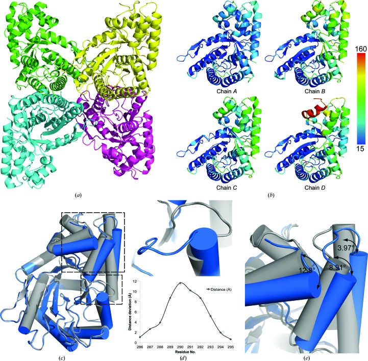
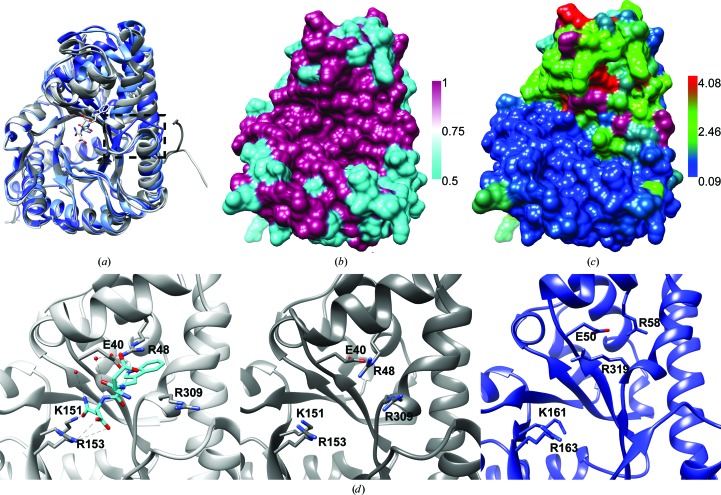
The apicomplexan parasite Toxoplasma gondii must invade host cells to continue its lifecycle. It invades different cell types using an actomyosin motor that is connected to extracellular adhesins via the bridging protein fructose-1,6-bisphosphate aldolase. During invasion, aldolase serves in the role of a structural bridging protein, as opposed to its normal enzymatic role in the glycolysis pathway. Crystal structures of the homologous Plasmodium falciparum fructose-1,6-bisphosphate aldolase have been described previously. Here, T. gondii fructose-1,6-bisphosphate aldolase has been crystallized in space group P22121, with the biologically relevant tetramer in the asymmetric unit, and the structure has been determined via molecular replacement to a resolution of 2.0 Å. An analysis of the quality of the model and of the differences between the four chains in the asymmetric unit and a comparison between the T. gondii and P. falciparum aldolase structures is presented.

Keywords: F16BP; MIC2; Toxoplasma; aldolase; glideosome; invasion.


Figures

Figure 1
Figure 1
(a) Cartoon representation of the TgAldolase tetramer with individually colored monomers. (b) Aligned chains colored by B factor in units of Å2. (c) Chain D (blue) aligned with chain A (gray) over the residue range 176–198 in the same orientation as the yellow chain in the tetramer above. (d) Depiction of the loop shift in chain D compared with chain A, with a plot representing the inter-residue Cα-atom deviation between residues 286 and 295. (e) Measurement of the degree of shift of α-helices 2, 11 and 12 between chains D and A. Figures were generated using PyMOL (Schrödinger).
Figure 2
Figure 2
2F oF c electron-density map (blue mesh) contoured at 1σ around residues 285–296 of chain A (left, gray) and chain D (right, blue), highlighting the different conformations of the loop connecting β-strand 10 to α-helix 10.
Figure 3
Figure 3
(a) Alignment via Chimera MatchMaker of PfAldolase (PDB entry 1a5c, chain A), PfAldolase bound to TRAP (PDB entry 2pc4, chain D) and TgAldolase (PDB entry 4tu1, chains A and D) represented as cartoons. The TRAP peptide shown in stick representation with two coordinating waters is present in the active site, which doubles as the adhesin tail binding pocket. A box highlights the dual conformation of the Met285–Ala296 loop in the TgAldolase chain A and D and PfAldolase structures. (b) Sequence conservation between TgAldolase and PfAldolase mapped on a surface representation of TgAldolase, with magenta indicating identical residues and cyan representing differences. (c) Surface representation of TgAldolase chain A colored according to residue r.m.s.d. (Å) from the reference structure PfAldolase (PDB entry 1a5c, chain A). (d) Three-panel enlargement of the adhesin-binding site of (left) TRAP bound to PfAldolase (PDB entry 2pc4), (middle) apo PfAldolase (PDB entry 1a5c) and (right) TgAldolase chain A, with residues important for TRAP binding highlighted as sticks.

References

    1. Adams, P. D. et al. (2010). Acta Cryst. D66, 213–221. - PubMed
    1. Bergman, L. W., Kaiser, K., Fujioka, H., Coppens, I., Daly, T. M., Fox, S., Matuschewski, K., Nussenzweig, V. & Kappe, S. H. (2003). J. Cell Sci. 116, 39–49. - PubMed
    1. Bosch, J., Buscaglia, C. A., Krumm, B., Ingason, B. P., Lucas, R., Roach, C., Cardozo, T., Nussenzweig, V. & Hol, W. G. J. (2007). Proc. Natl Acad. Sci. USA, 104, 7015–7020. - PMC - PubMed
    1. Buscaglia, C. A., Penesetti, D., Tao, M. & Nussenzweig, V. (2006). J. Biol. Chem. 281, 1324–1331. - PubMed
    1. Chen, V. B., Arendall, W. B., Headd, J. J., Keedy, D. A., Immormino, R. M., Kapral, G. J., Murray, L. W., Richardson, J. S. & Richardson, D. C. (2010). Acta Cryst. D66, 12–21. - PMC - PubMed

Publication types

Substances

Associated data