Review
. 2015 Jul:54:131-44.
doi: 10.1016/j.neubiorev.2014.08.013. Epub 2014 Sep 6.

Why do lesions in the rodent anterior thalamic nuclei cause such severe spatial deficits?

Affiliations
Review

Why do lesions in the rodent anterior thalamic nuclei cause such severe spatial deficits?

John P Aggleton et al. Neurosci Biobehav Rev. 2015 Jul.

Abstract

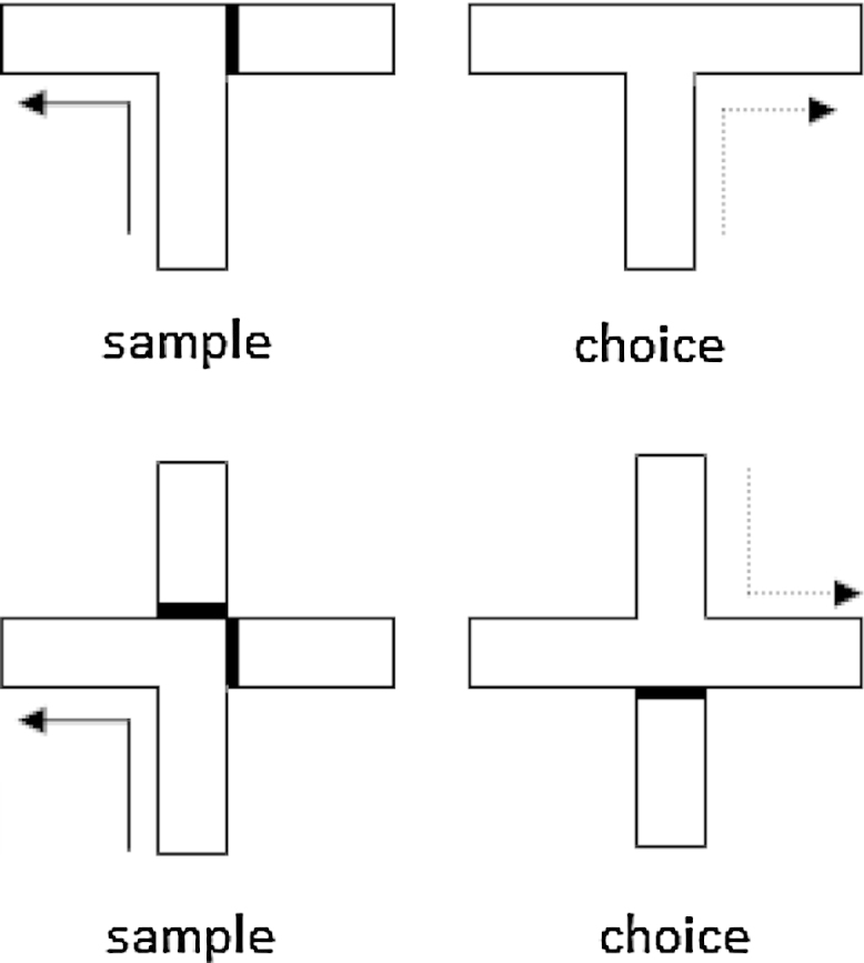
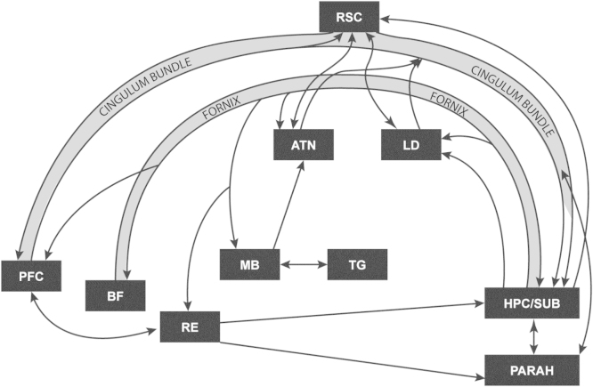
Lesions of the rodent anterior thalamic nuclei cause severe deficits to multiple spatial learning tasks. Possible explanations for these effects are examined, with particular reference to T-maze alternation. Anterior thalamic lesions not only impair allocentric place learning but also disrupt other spatial processes, including direction learning, path integration, and relative length discriminations, as well as aspects of nonspatial learning, e.g., temporal discriminations. Working memory tasks, such as T-maze alternation, appear particularly sensitive as they combine an array of these spatial and nonspatial demands. This sensitivity partly reflects the different functions supported by individual anterior thalamic nuclei, though it is argued that anterior thalamic lesion effects also arise from covert pathology in sites distal to the thalamus, most critically in the retrosplenial cortex and hippocampus. This two-level account, involving both local and distal lesion effects, explains the range and severity of the spatial deficits following anterior thalamic lesions. These findings highlight how the anterior thalamic nuclei form a key component in a series of interdependent systems that support multiple spatial functions.

Keywords: Alternation; Amnesia; Direction; Fornix; Learning; Mammillary bodies; Memory; Navigation; Space; Thalamus.


Figures

Fig. 1
Fig. 1
T-maze (upper) and cross-maze (lower) arrangements used to test spatial alternation. The bold line depicts the barrier used on sample runs (solid arrow) to control the arm choice by the animal. The dashed arrow shows the correct arm in the choice test. The cross-mazes show how opposing start positions can be used for the sample run and choice test in order to disrupt egocentric and directional alternation.
Fig. 2
Fig. 2
Schematic diagram illustrating how the rat hippocampal formation is associated with distinct sets of parallel, anterior thalamic connections. Connections conveyed via the fornix from the subiculum are shown with dashed lines. (Note that AD also receives nonfornical inputs from the postubiculum.) Double-headed arrows depict reciprocal connections. Abbreviations: DTG, dorsal tegmental nucleus of Gudden; MTT, mammillothalamic tract; VTGa, ventral tegmental nucleus of Gudden, pars anterior; VTGp, ventral tegmental nucleus of Gudden, pars posterior.
Fig. 3
Fig. 3
Performance of rats on reinforced T-maze alternation with lesions of the anterior thalamic nuclei (ANT) and closely related structures. The upper graph shows comparison performance (chance is 50%) from rats with fornix (FNX), mammillary body (MMB) and Sham lesions (Aggleton et al., 1995a,b), as well as rats with hippocampal (HPC) lesions (Aggleton et al., 1986). The lower graph compares the performance of rats with selective lesions centred in the anteromedial nucleus (AM), or the anteroventral and anterodorsal nuclei (AV/AD), as well as rats with combined lesions (ANT) involving all anterior thalamic nuclei (Aggleton et al., 1996). In all studies there was a retention interval of 10–15 s between the sample and choice run of each trial. The inter-trial interval was ∼4 min.
Fig. 4
Fig. 4
Schematic diagram showing some of the interconnections between sites implicated in spatial learning in rodents and anterograde amnesia in humans. Abbreviations: ATN—anterior thalamic nuclei; BF—basal forebrain (including septum and diagonal band); HPC/SUB—hippocampal formation (including subiculum); LD—laterodorsal thalamic nucleus; MB—mammillary bodies; PARAH—parahippocampal region; PFC—prefrontal cortex; RE—nucleus reuniens of the thalamus; RSC—retrosplenial cortex; TG—tegmental nucleus of Gudden.
Fig. 5
Fig. 5
Photomicrographs of brightfield coronal sections showing the extent of Fos protein staining (dark cells) in the retrosplenial cortex in rats with anterior thalamic lesions (ATN) or sham surgeries. The sections are taken from two subareas within the granular retrosplenial cortex (Rga and Rgb). The arrows highlight the dense Fos-positive staining in layer II in the sham controls, which contrasts with the marked depletion of Fos in the same layer in the rats with anterior thalamic lesions. The absence of Fos staining contrasts with the fact that the neurons are still present (e.g., when visualised with Nissl or NeuN staining).

References

    1. Aggleton J.P., Sahgal A. The contribution of the anterior thalamic nuclei to anterograde amnesia. Neuropsychologia. 1993;31:1001–1019. - PubMed
    1. Aggleton J.P., Hunt P.R., Rawlins J.N. The effects of hippocampal lesions upon spatial and non-spatial tests of working memory. Behav. Brain Res. 1986;19:133–146. - PubMed
    1. Aggleton J.P., Hunt P.R., Shaw C. The effects of mammillary body and combined amygdalar-fornix lesions on tests of delayed non-matching-to-sample in the rat. Behav. Brain Res. 1990;15:145–157. - PubMed
    1. Aggleton J.P., Keith A.B., Sahgal A. Both fornix and anterior thalamic, but not mammillary, lesions disrupt delayed nonmatching-to-position memory in rats. Behav. Brain Res. 1991;44:151–161. - PubMed
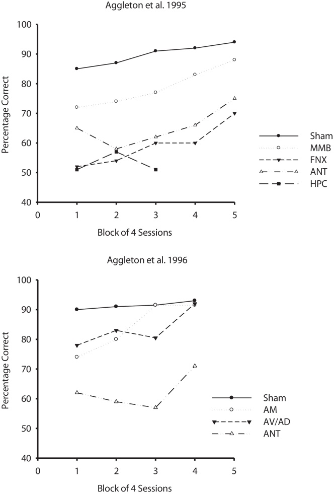
    1. Aggleton J.P., Neave N., Nagle S., Hunt P.R. A comparison of the effects of anterior thalamic, mamillary body and fornix lesions on reinforced spatial alternation. Behav. Brain Res. 1995;68:91–101. - PubMed

Publication types

Substances