

Quantification of left atrial strain and strain rate using Cardiovascular Magnetic Resonance myocardial feature tracking: a feasibility study
- PMID: 25196447
- PMCID: PMC4422260
- DOI: 10.1186/s12968-014-0060-6
Quantification of left atrial strain and strain rate using Cardiovascular Magnetic Resonance myocardial feature tracking: a feasibility study
Abstract
Background: Cardiovascular Magnetic Resonance myocardial feature tracking (CMR-FT) is a quantitative technique tracking tissue voxel motion on standard steady-state free precession (SSFP) cine images to assess ventricular myocardial deformation. The importance of left atrial (LA) deformation assessment is increasingly recognized and can be assessed with echocardiographic speckle tracking. However atrial deformation quantification has never previously been demonstrated with CMR. We sought to determine the feasibility and reproducibility of CMR-FT for quantitative derivation of LA strain and strain rate (SR) myocardial mechanics.
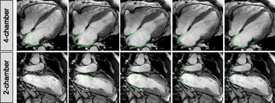
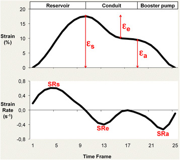
Methods: 10 healthy volunteers, 10 patients with hypertrophic cardiomyopathy (HCM) and 10 patients with heart failure and preserved ejection fraction (HFpEF) were studied at 1.5 Tesla. LA longitudinal strain and SR parameters were derived from SSFP cine images using dedicated CMR-FT software (2D CPA MR, TomTec, Germany). LA performance was analyzed using 4- and 2-chamber views including LA reservoir function (total strain [εs], peak positive SR [SRs]), LA conduit function (passive strain [εe], peak early negative SR [SRe]) and LA booster pump function (active strain [εa], late peak negative SR [SRa]).
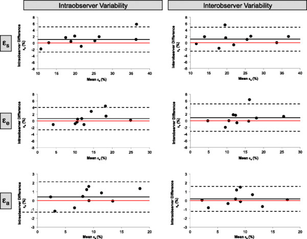
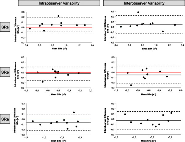
Results: In all subjects LA strain and SR parameters could be derived from SSFP images. There was impaired LA reservoir function in HCM and HFpEF (εs [%]: HCM 22.1 ± 5.5, HFpEF 16.3 ± 5.8, Controls 29.1 ± 5.3, p < 0.01; SRs [s⁻¹]: HCM 0.9 ± 0.2, HFpEF 0.8 ± 0.3, Controls 1.1 ± 0.2, p < 0.05) and impaired LA conduit function as compared to healthy controls (εe [%]: HCM 10.4 ± 3.9, HFpEF 11.9 ± 4.0, Controls 21.3 ± 5.1, p < 0.001; SRe [s]⁻¹: HCM -0.5 ± 0.2, HFpEF -0.6 ± 0.1, Controls -1.0 ± 0.3, p < 0.01). LA booster pump function was increased in HCM while decreased in HFpEF (εa [%]: HCM 11.7 ± 4.0, HFpEF 4.5 ± 2.9, Controls 7.8 ± 2.5, p < 0.01; SRa [s⁻¹]: HCM -1.2 ± 0.4, HFpEF -0.5 ± 0.2, Controls -0.9 ± 0.3, p < 0.01). Observer variability was excellent for all strain and SR parameters on an intra- and inter-observer level as determined by Bland-Altman, coefficient of variation and intraclass correlation coefficient analyses.
Conclusions: CMR-FT based atrial performance analysis reliably quantifies LA longitudinal strain and SR from standard SSFP cine images and discriminates between patients with impaired left ventricular relaxation and healthy controls. CMR-FT derived atrial deformation quantification seems a promising novel approach for the study of atrial performance and physiology in health and disease states.
Figures
References
-
- Kowallick JT, Edelmann F, Lotz J, Lamata P, Schuster A. Imaging diastolic dysfunction with cardiovascular magnetic resonance. J Cardiol Ther. 2014;1:58–64.
-
- Schuster A, Kutty S, Padiyath A, Parish V, Gribben P, Danford DA, Makowski MR, Bigalke B, Beerbaum P, Nagel E. Cardiovascular magnetic resonance myocardial feature tracking detects quantitative wall motion during dobutamine stress. J Cardiovasc Magn Reson. 2011;13:58. doi: 10.1186/1532-429X-13-58. - DOI - PMC - PubMed
Publication types
MeSH terms
Grants and funding
LinkOut - more resources
Full Text Sources
Other Literature Sources
Research Materials
