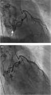
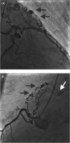
Unilateral and multilateral congenital coronary-pulmonary fistulas in adults: clinical presentation, diagnostic modalities, and management with a brief review of the literature
- PMID: 25196980
- PMCID: PMC6649402
- DOI: 10.1002/clc.22297
Unilateral and multilateral congenital coronary-pulmonary fistulas in adults: clinical presentation, diagnostic modalities, and management with a brief review of the literature
Abstract
Background: Congenital coronary-pulmonary fistulas (CPFs) are commonly unilateral, but bilateral and multilateral fistulas may occur. In multilateral CPFs, the value of a multidetector computed tomography (MDCT) imaging technique as an adjuvant to coronary angiography (CAG) is eminent. The purpose of this study was to describe the clinical presentation, diagnostic modalities, and management of coincidentally detected congenital CPFs.
Hypothesis: Unilateral and multilateral coronary-pulmonary fistulas are increasingly detected due to the wide speard application of multidetector computed tomography which might be a supplementary or replacing to conventional coronary angiography.
Methods: We evaluated 14 adult patients with congenital coronary artery fistulas (CAFs) who were identified from several Dutch cardiology departments.
Results: Fourteen adult patients (5 female and 9 male), with a mean age of 57.5 years (range, 24-80 years) had the following abnormal findings: audible systolic cardiac murmur (n = 4), chronic atrial fibrillation (n = 2), nonsustained ventricular tachycardia (n = 1), and cardiomegaly on chest x-ray (n = 2). Echocardiography revealed normal findings with trivial valvular abnormalities (n = 9), depressed left ventricle systolic function (n = 3), and severe mitral regurgitation and atrial dilatation (n = 2). The findings in the rest of the patients were unremarkable. CAG and MDCT were used as a diagnostic imaging techniques either alone (CAG, n = 6; MDCT, n = 1) or in combination (n = 7). Single modality and multimodality diagnostic methods revealed 22 fistulas including CPFs (n = 15), coronary cameral fistulas terminating into the right (n = 2) and the left atrium (n = 1), and systemic-pulmonary fistulas (n = 4). Of all of the fistulas, 10 were unilateral, 6 were bilateral, and 6 was hexalateral. (13) N-ammonia positron emission tomography-computed tomography was performed in 3 patients revealing decreased myocardial perfusion reserve.
Conclusions: CAG remains the gold standard for detection of CPFs. An adjuvant technique using MDCT provides full anatomical details of the fistulas.
© 2014 Wiley Periodicals, Inc.
Figures


References
-
- Krause W. Uber der ursprung einer akzessorische A coronaria aus der A pulmonalis. Z Rationell Med. 1865;24:225–227.
-
- Hagau AM, Dudea SM, Hagau R, et al. Large coronary pseudoaneurysm with pulmonary artery fistula, six months after left main trunk stenting with paclitaxel‐eluting stent. Med Ultrason. 2013;15:59–62. - PubMed
-
- Wei J, Azarbal B, Singh S, et al. Frequency of coronary artery fistulae is increased after orthotopic heart transplantation. J Heart Lung Transplant. 2013;32:744–746. - PubMed
-
- Musleh G, Jalal A, Deiraniya AK. Post‐coronary artery bypass grafting left internal mammary artery to pulmonary artery fistula: a 6 year follow‐up following successful surgical division. Eur J Cardiothorac Surg. 2001;20:1258–1260. - PubMed
-
- Bulur S, Satilmisoglu MH, Ozhan H, et al. Coronary‐to‐pulmonary artery fistula due to a penetrating trauma. Turk Kardiyol Dern Ars. 2012;40:109. - PubMed
Publication types
MeSH terms
LinkOut - more resources
Full Text Sources
Other Literature Sources

