

Prethreshold retinopathy in premature infants with intrauterine growth restriction
- PMID: 25196981
- PMCID: PMC6172005
- DOI: 10.1111/apa.12799
Prethreshold retinopathy in premature infants with intrauterine growth restriction
Abstract
Aim: To determine, among very preterm newborns, whether those who are growth-restricted are at increased risk of retinopathy of prematurity (ROP), and to explore whether the mixed findings of prior studies are the consequence of sampling based upon birthweight instead of gestational age.
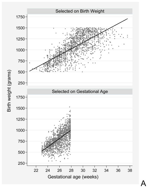
Methods: Using data from the ELGAN Study, we created logistic regression models of prethreshold ROP risk to adjust for confounders and calculate odds ratios and 99% confidence intervals. We created scatter plots to display the gestational age/birthweight relationship in infants enrolled in studies with different selection criteria.
Results: Low gestational age [23-24 weeks, OR 11.6 (2.9, 47); 25-26 weeks, 8.1 (2.1, 32)] and severe growth restriction [birthweight Z-score <-2, OR 9.1 (1.1, 76)] were associated with increased risk of prethreshold ROP. We documented in scatter plots that a sample defined by birthweight has an excess of gestationally older, severely growth-restricted newborns.
Conclusion: In this sample, low gestational age and severe growth restriction were associated with increased risk of prethreshold ROP.
Keywords: Extremely low gestational age newborn; Growth restriction; Prematurity; Retinopathy; Retinopathy of prematurity.
©2014 Foundation Acta Paediatrica. Published by John Wiley & Sons Ltd.
Figures
Comment in
-
Gestational age, birthweight, and their influence on neonatal outcome.Acta Paediatr. 2015 Jan;104(1):5-6. doi: 10.1111/apa.12818. Acta Paediatr. 2015. PMID: 25545224 No abstract available.
References
-
- Holmstrom G, Broberger U, Thomassen P. Neonatal risk factors for retinopathy of prematurity--a population-based study. Acta ophthalmologica Scandinavica. 1998;76:204–7. [Research Support, Non-U.S Gov’t] - PubMed
-
- Seiberth V, Linderkamp O. Risk factors in retinopathy of prematurity. a multivariate statistical analysis. Ophthalmologica Journal international d’ophtalmologie International journal of ophthalmology Zeitschrift fur Augenheilkunde [Comparative Study] 2000;214:131–5. - PubMed
-
- Allvin K, Hellstrom A, Dahlgren J, Gronlund MA. Birth weight is the most important predictor of abnormal retinal vascularisation in moderately preterm infants. Acta Paediatr. 2014;103:594–600. - PubMed
-
- Fortes Filho JB, Valiatti FB, Eckert GU, Costa MC, Silveira RC, Procianoy RS. Is being small for gestational age a risk factor for retinopathy of prematurity? A study with 345 very low birth weight preterm infants. J Pediatr (Rio J) 2009;85:48–54. [Comparative Study] - PubMed
-
- Bardin C, Zelkowitz P, Papageorgiou A. Outcome of small-for-gestational age and appropriate-for-gestational age infants born before 27 weeks of gestation. Pediatrics. 1997;100(2):E4. - PubMed
Publication types
MeSH terms
Grants and funding
LinkOut - more resources
Full Text Sources
Other Literature Sources
Medical
