. 2014 Sep 16;111(37):E3845-52.
doi: 10.1073/pnas.1414545111. Epub 2014 Sep 2.

Structural insight into the molecular mechanism of allosteric activation of human cystathionine β-synthase by S-adenosylmethionine

Affiliations

Structural insight into the molecular mechanism of allosteric activation of human cystathionine β-synthase by S-adenosylmethionine

June Ereño-Orbea et al. Proc Natl Acad Sci U S A. .

Abstract

Cystathionine β-synthase (CBS) is a heme-dependent and pyridoxal-5'-phosphate-dependent protein that controls the flux of sulfur from methionine to cysteine, a precursor of glutathione, taurine, and H2S. Deficiency of CBS activity causes homocystinuria, the most frequent disorder of sulfur amino acid metabolism. In contrast to CBSs from lower organisms, human CBS (hCBS) is allosterically activated by S-adenosylmethionine (AdoMet), which binds to the regulatory domain and triggers a conformational change that allows the protein to progress from the basal toward the activated state. The structural basis of the underlying molecular mechanism has remained elusive so far. Here, we present the structure of hCBS with bound AdoMet, revealing the activated conformation of the human enzyme. Binding of AdoMet triggers a conformational change in the Bateman module of the regulatory domain that favors its association with a Bateman module of the complementary subunit to form an antiparallel CBS module. Such an arrangement is very similar to that found in the constitutively activated insect CBS. In the presence of AdoMet, the autoinhibition exerted by the regulatory region is eliminated, allowing for improved access of substrates to the catalytic pocket. Based on the availability of both the basal and the activated structures, we discuss the mechanism of hCBS activation by AdoMet and the properties of the AdoMet binding site, as well as the responsiveness of the enzyme to its allosteric regulator. The structure described herein paves the way for the rational design of compounds modulating hCBS activity and thus transsulfuration, redox status, and H2S biogenesis.

Keywords: CBS domain; hydrogen sulfide.


Conflict of interest statement

The authors declare no conflict of interest.

Figures

Fig. 1.
Fig. 1.
Interactions between protein domains in basal hCBS. (A) In hCBSΔ516–525, residues Y484, N463, and S466 anchor the Bateman module (blue) to the protein core (gray) through H-bonds with the residues E201 and D198 from the loop L191–202, thus occluding the entrance to the catalytic pocket. (B) The CBS-specific activity of selected hCBS variants in the absence (blue bars) and the presence (red bars) of 300 µM AdoMet. hCBS enzyme species marked with “Δ” lack residues 516–525 and form dimers.
Fig. 2.
Fig. 2.
The 3D structure of hCBS and dCBS. Crystal structure of (A) hCBSΔ516–525, (B) dCBS, and (C) AdoMet-bound hCBS E201S. Similarly to dCBS, hCBS E201S associates in dimers in which the Bateman module from two subunits interacts to form an antiparallel CBS module. In hCBS E201S, the CBS module hosts two molecules of AdoMet. The slightly tilted orientation (∼40 degrees) of the regulatory domain with respect to the catalytic core is due to the presence of a symmetry-related molecule in the crystal (Fig. S3). (D) The high structural similarity existing between the CBS modules of dCBS and hCBS E201S (Fig. S2), allowed us to easily reorient the tilted CBS module to its most probable position in solution using the dCBS structure as template. The dash-dotted lines represent the planes containing the flat disk-like CBS modules in each protein.
Fig. 3.
Fig. 3.
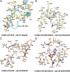
AdoMet binding site in basal and activated hCBS. (A) Site S1 in basal hCBSΔ516–525. The entrance to cavity S1 is sterically occluded by the presence of structural elements from the catalytic core of a complementary monomer in the dimer (cyan). Additionally, bulky hydrophobic residues occupy the cleft and impede the binding of AdoMet at this site. (B) Site S1 in activated AdoMet-bound hCBS E201S. Despite the presence of AdoMet during the crystallization, site S1 remained empty in our crystals. As shown, binding of AdoMet (in black lines) at site S1 would cause steric clashes within the cavity S1, even in the activated conformation of hCBS. Note: The potential location of AdoMet at site S1 has been modeled by performing a structural alignment of site S2 and S1, both in their activated conformations. (C) Site S2 in basal hCBSΔ516–525. The S2 cavity is fully solvent-exposed and is not blocked by bulky residues. In hCBS, AdoMet binds at a previously proposed site S2 of the Bateman module and induces a relative rotation of the two CBS motifs that results in a slight reorientation of the residues within the S2 cavity. In the absence of such structural change, accommodation of AdoMet within site S2 would be sterically impeded. (D) Site S2 in the activated AdoMet-bound hCBS E201S. This cavity represents the unique AdoMet binding site. The S2 cavity shows a hydrophobic cage that hosts the adenine ring of AdoMet, conserved aspartate (D538), threonine (T535), and serine (S420) residues to stabilize the ribose ring, and a hydrophobic residue (I537) preceding D538 that accommodates the alkyl chain of AdoMet.
Fig. 4.
Fig. 4.
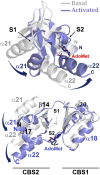
Effect of AdoMet on the Bateman module of hCBS. Two different views of the structural superimposition of the Bateman modules of hCBSΔ516–525 (gray; PDB ID code 4L3V) and hCBS E201S (blue; PDB ID code 4PCU). The latter contains one molecule of AdoMet (sticks) bound at site S2. AdoMet induces a conformational change in the Bateman module that consists of a relative rotation of the two CBS motifs without altering their overall secondary and tertiary structure. The root-mean-square deviations (rmsd) between the CBS1-CBS1* and CBS2-CBS2* motifs of both proteins are 1.211 and 1.017, respectively. The large structural difference between Bateman modules in the basal and in the activated states is reflected in an rmsd of 2.647. Blue arrows indicate the direction of the rotation upon AdoMet binding. The location of sites S1 and S2 is also indicated.
Fig. 5.
Fig. 5.
Interactions that stabilize the activated conformation of CBSs. Several hydrophobic interactions between helices α12 (core) and α15 (connective linker) stabilize the orientation of the helix α15 in both dCBS (A) and AdoMet-bound hCBS E201S (C). (B) Network of polar interactions involving the residues R310, D362, N363, E366, K371, R475, and E478 determine in dCBS the orientation of the α-helices equivalent to α12, α15, and α21 of hCBS. (D) On the other hand, in AdoMet-bound hCBS E201S, the presence of residues L392, L397, and H501 (in equivalent positions to E366, K371, and R475, respectively, in dCBS) weakens the polar network between helices α15 and α21 and presumably facilitates hCBS activation and the displacement of the Bateman module. In the hCBS E201S crystals, the region corresponding to residues 398–405 of hCBS (dark gray) is disordered. To facilitate the structural comparison, this region has been modeled using the dCBS structure as template. The side chains of residues R389 and H501 are not represented because their orientation is not clear in the electron density maps. (E) Sequence alignment of dCBS and hCBS in these regions.
Fig. 6.
Fig. 6.
A model summarizing the effect of AdoMet binding and/or pathogenic mutations on the structure of hCBS. The hCBSΔ516–525 associates in dimers (the subunits are depicted in orange and blue, respectively). AdoMet binding triggers a conformational change that makes the protein to progress from a basal (A) toward an activated (B) state. In addition, the pathogenic mutation, such as D444N, may partially activate the enzyme by slightly modifying the dimer structure that still retains the overall fold of the basal conformation (C). Under these circumstances, the loops delineating the entrance of the catalytic cavity (represented by red bars) find some more space to move, thus contributing to increase in the basal activity of the enzyme. The progression of these mutants toward the activated state (B) in the presence of AdoMet depends on the effect of a particular mutation on the formation and/or stability of the CBS module. (D) Other mutations, such as the S466L or the artificial E201S, which impair the interactions between the Bateman module and the catalytic core (as illustrated in Fig. 1A), promote a more significant displacement of the regulatory domain away from the catalytic cavity, without formation of the disk-like CBS module. If AdoMet binding is not impaired, these mutants can easily progress toward the activated conformation as found in hCBS E201S in the presence of AdoMet (B). Both states depicted in B and D allow for free movement of the entrance loops and subsequently unrestricted flow of substrates into the PLP-containing active site.

References

    1. Miles EW, Kraus JP. Cystathionine beta-synthase: Structure, function, regulation, and location of homocystinuria-causing mutations. J Biol Chem. 2004;279(29):29871–29874. - PubMed
    1. Mudd SH, Levy HL, Kraus JP. Disorders of transsulfuration. In: Scriver CR, et al., editors. The Metabolic and Molecular Bases of Inherited Disease. 8th Ed. New York: McGraw-Hill; 2001. pp. 2007–2056.
    1. Banerjee R, Evande R, Kabil O, Ojha S, Taoka S. Reaction mechanism and regulation of cystathionine beta-synthase. Biochim Biophys Acta. 2003;1647(1-2):30–35. - PubMed
    1. Majtan T, Singh LR, Wang L, Kruger WD, Kraus JP. Active cystathionine beta-synthase can be expressed in heme-free systems in the presence of metal-substituted porphyrins or a chemical chaperone. J Biol Chem. 2008;283(50):34588–34595. - PMC - PubMed
    1. Christen P, Mehta PK. From cofactor to enzymes: The molecular evolution of pyridoxal-5′-phosphate-dependent enzymes. Chem Rec. 2001;1(6):436–447. - PubMed

Publication types

Substances

Associated data